


Application of ex vivo liver perfusion in hepatoma research
 0
0 Abstract
Hepatocellular carcinoma remains a leading cause of cancer-related mortality worldwide. Despite advances in surgical and systemic therapies, recurrence rates remain high, and translational models for therapeutic testing are limited. This review explores the evolving role of ex vivo liver perfusion (EVLP) as a translational platform in hepatoma research, highlighting its applications in tumour modelling, therapeutic testing, and biomarker discovery. A narrative synthesis of recent literature was performed, focusing on EVLP modalities such as normothermic machine perfusion, hypothermic oxygenated perfusion, split-liver perfusion, and segmental perfusion of resected tumour-bearing tissue. EVLP preserves hepatic architecture and metabolic function, enabling real-time study of tumour microenvironments, pharmacological responses, and recurrence mechanisms. Segmental perfusion provides an ethically viable translational model. Overall, EVLP represents a transformative tool in hepatobiliary oncology, bridging the gap between in vitro models and clinical practice, enhancing mechanistic understanding, and accelerating therapeutic innovation.
Keywords
INTRODUCTION
Management of primary liver cancer (PLC) and metastatic liver disease has attracted increasing attention in recent years due to the high mortality rate of PLC[1] and the frequent metastasis of colorectal cancer to the liver[2]. Globally, PLC has the 6th highest incidence and is the 3rd leading cause of cancer deaths among men and women, with approximately 80% of cases being hepatocellular carcinoma (HCC)[1]. Colorectal cancer ranks 3rd in incidence and 2nd in global mortality[1], with colorectal liver metastasis (CRLM) developing in the majority of patients with stage IV disease[3]. Although advancements in neoadjuvant chemotherapy and surgical techniques have improved outcomes for CRLM[4,5], HCC continues to present significant challenges due to high recurrence rates and treatment resistance. Liver transplantation and resection remain curative options; however, recurrence rates post-transplantation have been estimated at 15%-19% in selected cohorts[6]. Traditional in vitro models fail to replicate the complexity of the tumour microenvironment (TME), while in vivo studies are limited by ethical and logistical constraints.
Following the first use of ex vivo organ perfusion (EVOP) in 1935[7], EVOP has mainly been utilised in the field of organ transplantation and, in the context of ex vivo liver perfusion (EVLP) with the aim to expand the availability of donor organs[8]. Alternative areas of research other than transplantation have also been investigated and this review will discuss how EVLP has been developed predominantly for transplantation, but how these modalities are also applicable to Hepatoma research. The use of animal experimentation in EVLP has helped improve our understanding in areas such as disease modelling[9] and the evaluation of drug efficacy[10] and metabolism[11]. While smaller animal models provide more efficient forms of disease modelling and cost effectiveness, porcine models have a similar genome to that of humans as well as offering greater efficiency and practicality during EVLP[12].
EVLP utilising diseased human livers unsuitable for transplantation has emerged as a translational research platform that maintains tissue viability under near-physiological conditions[13], thus avoiding ethical concerns associated with animal models.
This review synthesises current applications of EVLP in hepatoma research and outlines future directions for its integration into experimental oncology.
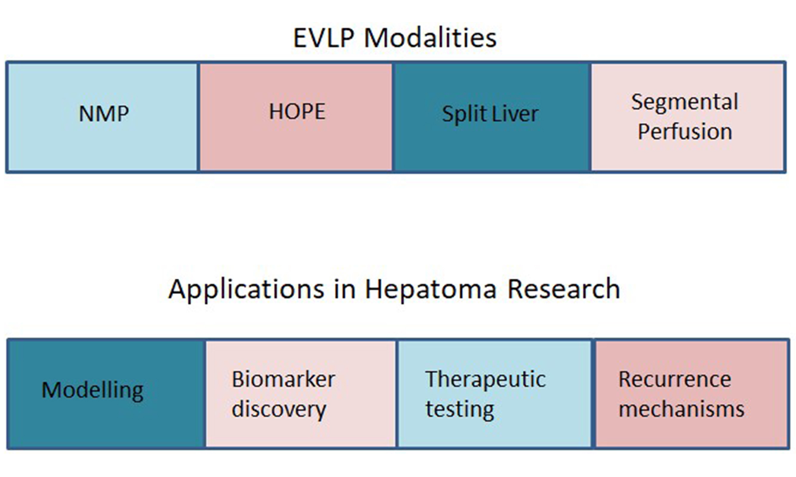
PRINCIPLES AND MODALITIES
EVLP involves the extracorporeal circulation of oxygenated perfusate through the hepatic vasculature. The two main modalities employed are normothermic machine perfusion (NMP) and hypothermic oxygenated perfusion (HOPE). In both approaches, the equipment and tissue typically include the hepatic specimen, the perfusion system, and auxiliary components. Given the liver’s dual blood supply, the portal vein (PV) is usually the afferent vessel, with the hepatic artery (HA) incorporated if required, and the hepatic veins serving as the efferent vessels. Once cannulated and connected, blood can be circulated in either pulsatile or continuous flow, depending on the pump type (centrifugal or roller). An oxygenator allows the blood to be re-oxygenated before returning to the liver. To maintain nutrition and metabolic support, the perfusate contains varying concentrations of constituents, such as glucose and vitamins, according to the specific protocol. In addition to controlling flow and pressure, the system temperature can be adjusted to achieve either physiological or hypothermic conditions.
An overview of the different EVLP modalities discussed herein is presented in Table 1.
Comparative overview of ex vivo liver perfusion modalities applied in hepatoma research
| Perfusion modality | Temperature | Tissue format | Applications in hepatoma research | Advantages | Limitations | Representative studies |
| NMP | 37 °C | Whole liver Split-liver Segmental | - Drug testing - TME modelling - Biomarker discovery - Gene therapy | - Preserves metabolic activity - Real-time functional assessment | - Complex setup - Higher cost | Nasralla et al.[14] Boteon et al.[18] Zimmerman and Carter[24] Kanani et al.[38] Trebo et al.[42] Brevini et al.[50] |
| HOPE | 4-10 °C | Whole liver | - Recurrence mechanism studies - Transplant oncology outcomes | - Reduces ischemia-reperfusion injury - Enhances graft viability | - Limited metabolic activity - Less suitable for drug testing | Ravaioli et al.[26] Schlegel et al.[28] Jeddou et al.[29] Mueller et al.[52] |
| Split-liver perfusion | Variable | Split lobes from donor liver | - Internal control for comparative studies - Tumour vs. non-tumour analysis | - Enhances experimental rigour - Reduces organ demand | -Requires suitable donor anatomy - Limited availability | Attard et al.[34] Lau et al.[36] Krendl et al.[43] Cabanes-Creus et al.[49] |
| Segmental perfusion | 37 °C | Resected tumour-bearing segments | - Ethical translational modelling - TME profiling | - Uses surgical waste tissue - High translational relevance | - Short perfusion duration - Limited tissue volume | Kanani et al.[38] Kanani et al.[39] An et al.[40] |
Normothermic perfusion
By maintaining the system at physiological temperature (37 °C), NMP aims to preserve resected livers in a near-physiological state. This enables the assessment of metabolic activity and hepatic function, histological changes, pharmacological responses, and immune profiling.
NMP has been shown to result in a lower proportion of graft injury compared to static cold storage (SCS) during transplantation[14]. The functionality of hepatic tissue can be assessed by monitoring bile production and the synthesis of coagulation factors during NMP[15]. In addition, Lascaris et al. evaluated metabolic activity by measuring concentrations of substrates in the exiting perfusate and collected histological samples after 72 h and 7 days[16]. These observations revealed mild ischaemic changes but preserved tissue architecture. Longer durations of NMP have also been demonstrated; Cillo et al. analysed a liver undergoing NMP for 424 h[17]. The specimen exhibited minimal cell death on days 13 and 15, and at the end of perfusion. Preserved metabolic and synthetic function was noted, highlighting the feasibility of long-term perfusion of resected livers.
Examples of pharmacological testing during NMP include analyses of lipid metabolism. Steatotic livers have been of particular interest in transplantation research due to the potential to expand the pool of extended criteria donors. This prompted studies investigating lipid metabolism in discarded human livers.
NMP has also enabled immune profiling of donor livers. Lee et al. and Hautz et al. demonstrated that cell lineages change during NMP, and the immunological response becomes less pro-inflammatory over time[20,21]. Further work by Hautz et al. aimed to identify genetic patterns predictive of graft viability[22]. They found multiple genes that may be useful in determining graft outcomes, including genes such as CD274 and liver enriched antimicrobial peptide 2 (LEAP2). Notably, programmed cell-death ligand 1 (PD-L1), encoded by CD274, is not only predictive of graft outcomes but has also been implicated in the prognosis of HCC[23].
Despite the significant research opportunities offered by NMP, the cost-effectiveness of NMP and hypothermic machine perfusion in the UK remains a crucial issue[24]. Zimmerman and Carter noted that cost-effectiveness should be re-evaluated once further long-term data on NMP and associated costs become available[24].
HOPE
HOPE operates at a lower temperature than NMP (usually 4-10 °C). Maspero et al. elucidate the pathways involved in ischaemia-reperfusion injury (IRI) and show that HOPE reduces the extent of IRI during transplantation and promotes regenerative mechanisms[25].
The single-centre randomised controlled trial (RCT) by Ravaioli et al. found that HOPE, following a period of SCS, reduced early graft dysfunction and resulted in a lower proportion of complications and re-admissions in the post-operative period compared to SCS alone[26]. Positive 5-year graft and patient survival outcomes have been reported in the multicentre, observational HOPE-REAL study (NCT05520320)[27], the largest study to date, with death-censored survival rates exceeding 80%. Schlegel et al. conducted a multicentre RCT comparing livers allocated to SCS alone or to SCS followed by HOPE[28]. While HOPE did not reduce the number of patients developing at least one major complication (defined as Clavien-Dindo score > III), it did reduce the number of patients developing serious liver-related complications (CD > IIIb).
A prominent issue highlighted in recent literature is the need for validated markers to assess graft viability during transplantation. Multiple markers and parameters have been investigated for their predictive value during NMP; however, no consensus exists, leading to discrepancies in parameters used to assess graft viability, as discussed by Jeddou et al.[29]. Jeddou et al. question whether grafts failing proposed criteria could function adequately if transplanted and advocate for graft scoring systems instead of binary parameters[29]. Although HOPE occurs at lower temperatures than NMP, reducing metabolism in perfused livers, a recent international observational study by Eden et al. demonstrated the potential for developing biomarkers of graft viability[30]. Flavin mononucleotide (FMN), a mitochondrial complex I cofactor, is released upon mitochondrial damage. Eden et al. show that FMN can be detected during dual HOPE (DHOPE, perfusion of both PV and HA) and has predictive value for graft viability[30]. As with NMP, no consensus exists on the optimal FMN levels to assess perfusate or judge graft viability.
The efficacy of NMP and HOPE has been established in comparison to SCS; however, few studies have directly compared NMP and HOPE. Lurje et al. demonstrated that immune cell populations differ between the two modalities, with smaller populations of leucocytes and granulocytes observed in the effluent of HOPE-treated livers[31]. They also found that PD-L1 expression on the cell surface differed depending on whether NMP or HOPE was utilised. The clinical relevance of this finding remains unclear, and further studies are required to assess its significance.
Split-liver perfusion and segmental perfusion
Following advancements with NMP and HOPE in transplantation, innovations such as split-liver and segmental perfusion using resected tumour-bearing livers have expanded the translational utility of EVLP, enabling ethical and reproducible experimentation on human livers.
Split-liver perfusion involves separating the two physiological lobes of the liver, which can then undergo machine perfusion. The advantages and limitations of NMP and HOPE in split-liver perfusion have been discussed, with themes similar to those observed in whole-liver perfusion[32,33]. Beyond transplantation, improvements in split-liver perfusion protocols may facilitate more in-depth preclinical testing of therapeutics, as the lobes exhibit near-equal metabolism and functionality[34] and can maintain long-term perfusion over multiple days[35]. Lau et al. further developed long-term split-liver perfusion, reporting two livers declined for transplantation that were split and maintained on NMP for over 12 days[36]. Throughout this period, metabolic and functional parameters remained stable, and tissue architecture was preserved. However, bacterial cultures of the perfusate were positive at 306 h, despite being negative at 96 h, suggesting that protocol optimisation is required to extend the safe duration of these studies[37].
Segmental liver perfusion is an exciting new area for research opportunities. Kanani et al. have demonstrated viability of hepatic cells in segmented diseased livers undergoing NMP[38]. Nine livers were resected due to HCC or colorectal liver metastases, with healthy segments being taken from each of the segments to be perfused. There was a comparison made between the perfusion of expired O negative red blood cells (RBCs) and Oxyglobin/Volplex. Following 6 h of NMP, 4 of 5 segments perfused with the oxyglobin solution demonstrated intact histology compared to 1 of 4 segments perfused with expired RBCs. Though the sample size used in this study was small, it highlights an exciting opportunity for further preclinical research and the development of longer-term models similar to those of whole or split-liver perfusion.
The potential use of segmental perfusion in the context of disease modelling is also evidenced by
APPLICATIONS IN HEPATOMA RESEARCH
TME modelling
Trebo et al. have conducted an impressive study utilising EVLP to model CRLM[42]. Six livers obtained from patients undergoing either resection or transplantation due to CRLM underwent NMP. Throughout the study (median duration of NMP was 41 h, with maximum duration of 144 h), tissue architecture of the livers was sustained as were the metabolic/functional activities of both healthy and tumoral tissue. Though not directly examined, implications of the constant perfusion pressure/flow during NMP imply that the vascular integrity of the specimens, on a macro/microscopic level, is maintained. An interesting area of further research would be to directly assess microvascular integrity during NMP. The population of immune cell-types present varied greatly between livers, with most of this variability arising not from healthy tissue, but from the cancerous environment. Trebo et al. also showed how immunosuppressive cell types play a role in the maintenance of the TME by creating a hypoxic and fibrotic environment in which the tumour cells can reside[42].
In the study by Trebo et al., assessment of tumour and healthy liver tissue was performed[42]. A slight, but important alternate view is shown by Krendl et al., in which 2 different pathologies were examined within the same liver[43]. By analysing a resected cirrhotic liver with HCC in situ, they were able to maintain NMP for 96 h, finding similar tissue viabilities and histological preservation in both tumour and cirrhotic biopsies. Though not directly assessing the TME, Krendl et al. explain how their study allows assessment of two different pathologies within the same specimen[43]. This hopefully will aid in the development of more in-depth studies of the TME of HCC, the ex vivo study of mechanisms involved in cirrhosis and the mechanisms leading to development of HCC in cirrhotic livers.
As an aside, given the difficulties in translational application of animal studies, microphysiological systems (MPSs) have been developed to attempt to bridge the gap of studying ex vivo models with near in vivo conditions at a cellular level. These MPSs use human cells to develop a micro-environment similar to that of in vivo situations, as discussed by Clark et al.[44]. This paper discusses the different MPSs available and how they provide an opportunity to study the beginnings of tumour progression/metastasis in the controlled micro-environment. Though MPSs hold promise in the establishment of early metastatic models, only one validated MPS exists for investigating metastases, and similar problems to those of EVLP remain, such as the translation of findings to in vivo conditions over longer periods.
Therapeutic testing, pharmacological profiling and genetic testing
There were no studies examining the role of oncological treatments during EVLP in this narrative review search. This is clearly an area of great potential going forward with future studies. Smaller-scale models have been able to culture cancerous hepatic tissue and interrogate them with anti-cancer therapies. Precision-cut tissue slicing (PCTS) allows the use of resected HCC livers for testing of immunotherapy and chemotherapy agents. After developing the cultured specimens, Jagatia et al. administered doxorubicin (a chemotherapeutic drug) and multiple immunotherapeutic drugs, including atezolizumab with bevacizumab (a combination often used for HCC treatment) to the PCTS[45]. Results were monitored over 8 days showing changes in the degree of proliferation and viability of tumour cells. Given the evidence for NMP and HOPE in the maintained viability of resected tissue over several days, there are numerous exciting avenues for further investigations. One avenue would obviously be the testing of novel chemotherapy/immunotherapy drugs in HCC/CRLM livers. Once administered, specimens could then be taken at different time points for analysis. With the use of split-liver perfusion, one lobe could be exposed to this drug as the experimental wing and be compared to the control lobe, aiming at minimising heterogeneity between livers. This would allow assessment of drug efficacy, the pharmacodynamics/kinetics of the compound, and potential resistance mechanisms. Given the advancements in prolonged EVLP (as previously discussed in this review), monitoring could be carried out over a longer term than that seen in smaller models, such as that reported by Jagatia et al.[45]. A second interesting area of further study would be to subject tumour preclinical locoregional therapy such as embolisation and ablative techniques.
EVLP has been used in the development of therapeutics aimed at reducing IRI. Nanoceria (NC) is a nanoparticle (NP) that acts as an antioxidant. Del Turco et al. administered NC to human livers with an endpoint objective to assess telomere attrition and mitochondrial DNA deletion (markers of DNA damage) compared to controls[46]. Liver used in this study underwent NMP, with five livers receiving NC and 4 controls receiving NMP alone. Del Turco et al. found that mitochondrial DNA deletion was significantly reduced in the experimental arm[46]. On the other hand, telomere attrition was not shown to be significantly reduced. Preclinical animal studies have produced encouraging findings in potential uses of NC discussed by Tang et al., but there is still limited evidence of NC use in humans[47]. Again, there is an exciting opportunity for further research into the role of NC in the context of HCC, given the link between IRI and HCC recurrence post-transplant[25].
In their review, Liu et al. discuss the current status of gene therapies to treat disease, including several ongoing clinical trials assessing the efficacy of gene-editing therapies[48]. In the context of EVLP and gene therapy, Cabanes-Creus et al. provide a proof-of-concept study utilising two discarded human livers commenced on NMP[49]. Following this, the livers were split and maintained on split-liver NMP, allowing comparison between control and experimental lobes. Multiple adeno-associated viral (AAV) vector variants were used during the study. To allow further comparisons, one of the livers used was perfused with anti-AAV antibodies, whilst the other was perfused with plasma containing low anti-AAV antibody levels. Cabanes-Creus et al. demonstrated that in the presence of circulating anti-AAV antibodies, the variant AAV8 was shown to have the highest functionality of those investigated based on vector clearance from perfusate, vector uptake and gene expression[49]. Further work utilising AAV8 has been carried out by
As yet, EVLP has not been routinely used for pharmacological profiling, but its potential for drug profiling has been demonstrated in a pilot study by Tingle et al.[51]. In this study, the drug 2,4-dinitrophenol (DNP) was administered to steatotic livers deemed unsuitable for transplantation and maintained using NMP. DNP is an oxidative phosphorylation uncoupler that was previously used as a weight-loss agent. A key manifestation of DNP toxicity is hyperthermia. Tingle et al. show that liver hyperthermia was transient and that the increase in oxygen consumption was dependent on DNP concentration[51]. Although this was a small study, it highlights the potential of EVLP for pharmacokinetic assessment.
Investigating recurrence mechanisms post-transplant
As previously discussed, recurrence rates of HCC can lie between 15%-19% in selected cohorts[6]. The association between IRI and tumour progression has led to multiple studies investigating whether the use of HOPE during liver transplantation has a protective effect in HCC recurrence. Mueller et al. compared the outcomes of livers transplanted having received HOPE perfusion, or not perfused at centre A[52]. Centre A included 70 livers that received HOPE following donation after circulatory death (DCD) and 70 unperfused livers following donation after brainstem death (DBD). In Centre B, outcomes of 70 unperfused DCD livers and 70 unperfused DBD livers were compared. In Centre A, unperfused livers were four times more likely to develop recurrence than HOPE-perfused livers. Patients receiving HOPE-perfused livers had a higher 5-year recurrence-free survival rate. Comparisons were also made between HOPE-perfused livers in Centre A and the unperfused DCD cohort in Centre B. Mueller et al. further demonstrated that unperfused DCD livers had a twofold higher recurrence rate than HOPE-perfused DCD livers[52].
Similarly favourable outcomes with HOPE have been reported by Dajti et al.[53]. In this retrospective cohort study, 60 livers underwent HOPE perfusion, whereas 177 received SCS. The risk of recurrence was lower and recurrence-free survival higher in HOPE-treated livers. In contrast, Rigo et al. observed no significant difference in recurrence rates between 80 DHOPE-treated livers (66 DBD, 14 DCD) and 246 SCS-treated livers[54]. These conflicting findings underscore the need for further studies to clarify the potential benefits of HOPE in reducing HCC recurrence.
The results of the HOPE4CANCER trial (NCT06717919) are eagerly anticipated. This ongoing, multicentre international RCT compares HOPE with SCS[55]. Livers in the study are allocated 1:1 to either cold storage alone or HOPE following cold storage, with recurrence-free survival as the primary endpoint.
Maspero et al. provide a review of HCC recurrence and IRI[25]. They discuss how recurrence following transplantation can be attributed to three main processes: (1) systemic release of cancer cells during surgery due to mechanical pressures experienced by the diseased liver; (2) mitochondrial damage during surgery leading to dysfunction and IRI; and (3) activation of innate hepatic repair mechanisms that may promote tumour recurrence.
Biomarker discovery and omics integration
EVLP enables serial sampling of perfusate and tissue, supporting identification of circulating tumour markers, transcriptomic and proteomic profiling, and metabolomic analysis. Integration with multi-omics platforms enhances mechanistic insights and supports precision oncology.
Hautz et al. use transcriptomic analysis to develop a predictive signature of graft quality[22]. During this analysis, they demonstrated alterations in gene expression, signalling pathways, and the dynamics of immune-parenchymal cell interactions. Although not directly related to transcriptomic analysis of HCC or CRLM, these findings demonstrate how NMP can be applied to transcriptomic profiling. In their comprehensive modelling of CRLM, Trebo et al. provide strong evidence of the interplay between multiple signalling pathways, diverse cellular phenotypes, and altered gene expression within the TME, and demonstrate that these features were preserved during NMP[42]. For example, C-X-C chemokine receptor type 4 (CXCR4) signalling, which is known to be involved in the migration of immunosuppressive cells and in various malignancies[56], showed increased activity within the TME.
Currently, radiological investigations [ultrasound scan (USS) and CT] and non-specific laboratory investigations [e.g., the serum protein alpha fetoprotein (AFP)] are used for the diagnosis and surveillance of HCC[57]. Although these investigations are useful, their low specificity and operational dependence limit their effectiveness, driving interest in the development of biomarkers and validated risk scores for HCC[58-60].
Though the studies discussed in this review show promising results, they involve varying, but generally small, sample sizes. As EVLP is increasingly used in clinical practice, results from future studies will provide stronger evidence and enable more targeted therapeutic studies using discarded human livers.
The long-term perfusion capabilities of EVLP, its ability to support multi-omic profiling, and the ongoing interest in sensitive and specific biomarkers provide promising avenues for future research. For example, a steatotic liver unsuitable for transplantation could be split and perfused. The experimental lobe could be exposed to an oncogenic stimulus, and the multi-omic profiles of the experimental and control lobes could be compared to identify novel biomarkers, uncover potential therapeutic targets, and further elucidate HCC pathophysiology in a human model.
ETHICAL BENEFITS OF EVLP
Historically, animal studies have been used to support the progression from preclinical studies to clinical trials and to improve understanding of disease concepts[63]. The 3Rs principle (replace, reduce, and refine) has been developed to optimise animal welfare and should be followed throughout animal experimentation. This principle is supported by international bodies, such as the European Medicines Agency (EMA)[64]. Although animal models have provided valuable insights into disease fundamentals[63], challenges remain in translating findings to humans and reproducing results in clinical trials[65]. These limitations have driven the development of alternatives, including in silico modelling and tissue engineering[66].
Although still relatively new, EVLP enables real-time study of human tissue obtained from patients undergoing resection or transplantation. This approach mitigates the ethical concerns associated with animal testing and overcomes the translational limitations inherent in animal models.
CONCLUSION
EVLP represents a transformative platform in hepatoma research. Its ability to preserve human liver tissue both functionally and structurally enables mechanistic studies, therapeutic development, and biomarker discovery. Segmental perfusion of resected tumour-bearing tissue provides an ethically viable model with strong translational relevance. As protocols mature and multi-omics integration progresses, EVLP is poised to redefine experimental standards in hepatobiliary oncology.
DECLARATIONS
Authors’ contribution
Data curation, writing - original draft: Brown B
Conceptualisation, methodology, supervision, review: Chung WY, Isherwood J
Availability of data and materials
Not applicable.
AI and AI-assisted tools statement
Not applicable.
Financial support and sponsorship
None.
Conflicts of interest
All authors declared that there are no conflicts of interest.
Ethical approval and consent to participate
Not applicable.
Consent for publication
Not applicable.
Copyright
© The Author(s) 2026.
REFERENCES
1. Bray F, Laversanne M, Sung H, et al. Global cancer statistics 2022: GLOBOCAN estimates of incidence and mortality worldwide for 36 cancers in 185 countries. CA Cancer J Clin. 2024;74:229-63.
2. Riihimäki M, Hemminki A, Sundquist J, Hemminki K. Patterns of metastasis in colon and rectal cancer. Sci Rep. 2016;6:29765.
3. Oki E, Ando K, Nakanishi R, et al. Recent advances in treatment for colorectal liver metastasis. Ann Gastroenterol Surg. 2018;2:167-75.
4. Adam R, Kitano Y. Multidisciplinary approach of liver metastases from colorectal cancer. Ann Gastroenterol Surg. 2019;3:50-6.
5. Tatsuta K, Sakata M, Kojima T, Booka E, Kurachi K, Takeuchi H. Updated insights into the impact of adjuvant chemotherapy on recurrence and survival after curative resection of liver or lung metastases in colorectal cancer: a rapid review and meta-analysis. World J Surg Oncol. 2025;23:56.
6. Bzeizi KI, Abdullah M, Vidyasagar K, Alqahthani SA, Broering D. Hepatocellular carcinoma recurrence and mortality rate post liver transplantation: meta-analysis and systematic review of real-world evidence. Cancers. 2022;14:5114.
8. Canizares S, Montalvan A, Chumdermpadetsuk R, Modest A, Eckhoff D, Lee DD. Liver machine perfusion technology: expanding the donor pool to improve access to liver transplantation. Am J Transplant. 2024;24:1664-74.
9. Krüger M, Ruppelt A, Kappler B, et al. Normothermic ex vivo liver platform using porcine slaughterhouse livers for disease modeling. Bioengineering. 2022;9:471.
10. Raigani S, Carroll C, Griffith S, et al. Improvement of steatotic rat liver function with a defatting cocktail during ex situ normothermic machine perfusion is not directly related to liver fat content. PLoS ONE. 2020;15:e0232886.
11. Stevens LJ, van de Steeg E, Doppenberg JB, Alwayn IPJ, Knibbe CAJ, Dubbeld J. Ex vivo gut-hepato-biliary organ perfusion model to characterize oral absorption, gut-wall metabolism, pre-systemic hepatic metabolism and biliary excretion; application to midazolam. Eur J Pharm Sci. 2024;196:106760.
12. Yeganeh M, Zito A, Sadat M, Pierro A, Rogers IM. Ex vivo organ perfusion systems for disease modeling and therapeutic applications in small animal models. J Tissue Eng Regen Med. 2025:16.
13. Liew B, Nasralla D, Iype S, Pollok JM, Davidson B, Raptis DA. Liver transplant outcomes after ex vivo machine perfusion: a meta-analysis. Br J Surg. 2021;108:1409-16.
14. Nasralla D, Coussios CC, Mergental H, et al.; Consortium for Organ Preservation in Europe. A randomized trial of normothermic preservation in liver transplantation. Nature. 2018;557:50-6.
15. Li J, Lu H, Zhang J, Li Y, Zhao Q. Comprehensive approach to assessment of liver viability during normothermic machine perfusion. J Clin Transl Hepatol. 2023;11:466-79.
16. Lascaris B, Woltjes LC, Bodewes SB, Porte RJ, de Meijer VE, Nijsten MWN. Metabolic balance of human livers during long-term normothermic machine perfusion. Am J Physiol Gastrointest Liver Physiol. 2025;328:G522-32.
17. Cillo U, Nalesso F, Bertacco A, Indraccolo S, Gringeri E. Normothermic perfusion of a human tumoral liver for 17 days with concomitant extracorporeal blood purification therapy: case description. J Hepatol. 2024;81:e96-8.
18. Boteon YL, Attard J, Boteon APCS, et al. Manipulation of lipid metabolism during normothermic machine perfusion: effect of defatting therapies on donor liver functional recovery. Liver Transpl. 2019;25:1007-22.
19. Nagrath D, Xu H, Tanimura Y, et al. Metabolic preconditioning of donor organs: defatting fatty livers by normothermic perfusion ex vivo. Metab Eng. 2009;11:274-83.
20. Lee ACH, Edobor A, Lysandrou M, et al. The effect of normothermic machine perfusion on the immune profile of donor liver. Front Immunol. 2022;13:788935.
21. Hautz T, Salcher S, Fodor M, et al. Immune cell dynamics deconvoluted by single-cell RNA sequencing in normothermic machine perfusion of the liver. Nat Commun. 2023;14:2285.
22. Hautz T, Hackl H, Gottschling H, et al. Transcriptomic signatures during normothermic liver machine perfusion correspond with graft quality and predict the early graft function. EBioMedicine. 2024;108:105330.
23. Zhang Y, Cui K, Yang Y, et al. Infiltration of a unique CD8+CD274+ cell subgroup in hepatocellular carcinoma is associated with poor clinical outcomes. J Hepatocell Carcinoma. 2023;10:1051-67.
24. Zimmermann J, Carter AW. Cost-utility analysis of normothermic and hypothermic ex-situ machine perfusion in liver transplantation. Br J Surg. 2022;109:e31-2.
25. Maspero M, Yilmaz S, Cazzaniga B, et al. The role of ischaemia-reperfusion injury and liver regeneration in hepatic tumour recurrence. JHEP Rep. 2023;5:100846.
26. Ravaioli M, Germinario G, Dajti G, et al. Hypothermic oxygenated perfusion in extended criteria donor liver transplantation-A randomized clinical trial. Am J Transplant. 2022;22:2401-8.
27. Eden J, Brüggenwirth IMA, Berlakovich G, et al. Long-term outcomes after hypothermic oxygenated machine perfusion and transplantation of 1,202 donor livers in a real-world setting (HOPE-REAL study). J Hepatol. 2025;82:97-106.
28. Schlegel A, Mueller M, Muller X, et al. A multicenter randomized-controlled trial of hypothermic oxygenated perfusion (HOPE) for human liver grafts before transplantation. J Hepatol. 2023;78:783-93.
29. Jeddou H, Tzedakis S, Chaouch MA, Sulpice L, Samson M, Boudjema K. Viability assessment during normothermic machine liver perfusion: a literature review. Liver Int. 2025;45:e16244.
30. Eden J, Thorne AM, Bodewes SB, et al. Assessment of liver graft quality during hypothermic oxygenated perfusion: the first international validation study. J Hepatol. 2025;82:523-34.
31. Lurje I, Uluk D, Hammerich L, Pratschke J, Tacke F, Lurje G. Comparing hypothermic oxygenated and normothermic liver machine perfusion: Translation matters. J Hepatol. 2024;80:e163-5.
32. Thorne AM, Lantinga V, Bodewes S, et al. Ex situ dual hypothermic oxygenated machine perfusion for human split liver transplantation. Transplant Direct. 2021;7:e666.
33. Lau NS, Ly M, Dennis C, et al. Liver splitting during normothermic machine perfusion: a novel method to combine the advantages of both in-situ and ex-vivo techniques. HPB. 2023;25:543-55.
34. Attard JA, Osei-Bordom DC, Boteon Y, et al. Ex situ normothermic split liver machine perfusion: protocol for robust comparative controls in liver function assessment suitable for evaluation of novel therapeutic interventions in the pre-clinical setting. Front Surg. 2021;8:627332.
35. Lau NS, Ly M, Dennis C, et al. Long-term ex situ normothermic perfusion of human split livers for more than 1 week. Nat Commun. 2023;14:4755.
36. Lau NS, Ly M, Dennis C, et al. Long-term normothermic perfusion of human livers for longer than 12 days. Artif Organs. 2022;46:2504-10.
37. Lau NS, Ly M, Dennis C, et al. Microbial contamination during long-term ex vivo normothermic machine perfusion of human livers. Transplantation. 2024;108:198-203.
38. Kanani T, Isherwood J, Chung WY, et al. A O03 ex vivo perfusion of isolated human liver segments: the development of a novel model for ethical, translational research. Br J Surg. 2022;109:znac404.003.
39. Kanani T, Alnabati N, Tang S, et al. HPB O04 segmental perfusion of ex-vivo human liver segments: a 3Rs compliant directly translatable model for pharmacological, bacteriological and genetic research. Br J Surg. 2023;110:znad348.018.
40. An H, Qian C, Huang Y, et al. Functional vulnerability of liver macrophages to capsules defines virulence of blood-borne bacteria. J Exp Med. 2022;219:e20212032.
41. Ferreira GS, Veening-Griffioen DH, Boon WPC, Moors EHM, van Meer PJK. Levelling the translational gap for animal to human efficacy data. Animals. 2020;10:1199.
42. Trebo M, Maurer T, Krendl FJ, et al. Ex vivo modelling of human colorectal cancer liver metastasis by normothermic machine perfusion. Mol Cancer. 2025;24:264.
43. Krendl FJ, Cardini B, Zoller H, Schneeberger S, Oberhuber R. Leveraging normothermic liver machine perfusion as a platform for oncologic assessment in cirrhotic livers. J Hepatol. 2025;82:e12-4.
44. Clark AM, Ma B, Taylor DL, Griffith L, Wells A. Liver metastases: microenvironments and ex-vivo models. Exp Biol Med. 2016;241:1639-52.
45. Jagatia R, Doornebal EJ, Rastovic U, et al. Patient-derived precision cut tissue slices from primary liver cancer as a potential platform for preclinical drug testing. EBioMedicine. 2023;97:104826.
46. Del Turco S, Cappello V, Tapeinos C, et al. Cerium oxide nanoparticles administration during machine perfusion of discarded human livers: a pilot study. Liver Transpl. 2022;28:1173-85.
47. Tang JLY, Moonshi SS, Ta HT. Nanoceria: an innovative strategy for cancer treatment. Cell Mol Life Sci. 2023;80:46.
48. Liu F, Li R, Zhu Z, Yang Y, Lu F. Current developments of gene therapy in human diseases. MedComm. 2024;5:e645.
49. Cabanes-Creus M, Liao SHY, Gale Navarro R, et al. Harnessing whole human liver ex situ normothermic perfusion for preclinical AAV vector evaluation. Nat Commun. 2024;15:1876.
50. Brevini T, Swift L, Reynolds H, et al. Successful AAV8 gene therapy on hepatic ex situ machine perfusion for mitochondrial neurogastrointestinal encephalomyopathy. J Hepatol. 2025;83:1218-25.
51. Tingle SJ, Thompson ER, Bates L, et al. Pharmacological testing of therapeutics using normothermic machine perfusion: a pilot study of 2,4-dinitrophenol delivery to steatotic human livers. Artif Organs. 2022;46:2201-14.
52. Mueller M, Kalisvaart M, O’Rourke J, et al. Hypothermic oxygenated liver perfusion (HOPE) prevents tumor recurrence in liver transplantation from donation after circulatory death. Ann Surg. 2020;272:759-65.
53. Dajti G, Germinario G, Prosperi E, et al. The role of cold ischemia time and hypothermic perfusion in predicting early hepatocellular carcinoma recurrences after liver transplantation. Artif Organs. 2024;48:619-25.
54. Rigo F, De Stefano N, Patrono D, et al. Impact of hypothermic oxygenated machine perfusion on hepatocellular carcinoma recurrence after liver transplantation. J Pers Med. 2023;13:703.
55. Eden J, Müller PC, Kuemmerli C, et al.; HOPE4Cancer Trial Investigators. Hypothermic oxygenated perfusion (HOPE) against cancer recurrence after liver transplantation for hepatocellular carcinoma-study protocol for an international multicenter randomized controlled trial (HOPE4Cancer). Trials. 2025;26:369.
56. Bianchi ME, Mezzapelle R. The chemokine receptor CXCR4 in cell proliferation and tissue regeneration. Front Immunol. 2020;11:2109.
57. Zhao J, Hu Z, Zheng X, et al. Blood biomarkers of hepatocellular carcinoma: a critical review. Front Cell Dev Biol. 2024;12:1489836.
58. Fan R, Papatheodoridis G, Sun J, et al. aMAP risk score predicts hepatocellular carcinoma development in patients with chronic hepatitis. J Hepatol. 2020;73:1368-78.
59. Hao X, Fan R, Zeng HM, Hou JL. Hepatocellular carcinoma risk scores from modeling to real clinical practice in areas highly endemic for hepatitis B infection. J Clin Transl Hepatol. 2023;11:1508-19.
60. Liu Z, Yuan H, Suo C, et al. Point-based risk score for the risk stratification and prediction of hepatocellular carcinoma: a population-based random survival forest modeling study. EClinicalMedicine. 2024;75:102796.
61. Barjasteh AH, Jaseb Mazhar AleKassar R, Al-Asady AM, et al. Therapeutic potentials of miRNA for colorectal cancer liver metastasis treatment: a narrative review. Iran J Med Sci. 2025;50:202-19.
62. Amr KS, Elmawgoud Atia HA, Elazeem Elbnhawy RA, Ezzat WM. Early diagnostic evaluation of miR-122 and miR-224 as biomarkers for hepatocellular carcinoma. Genes Dis. 2017;4:215-21.
63. Domínguez-Oliva A, Hernández-Ávalos I, Martínez-Burnes J, Olmos-Hernández A, Verduzco-Mendoza A, Mota-Rojas D. The importance of animal models in biomedical research: current insights and applications. Animals. 2023;13:1223.
64. EMA. Regulatory acceptance of 3R (replacement, reduction, refinement) testing approaches - Scientific guideline. Available from: https://www.ema.europa.eu/en/regulatory-acceptance-3r-replacement-reduction-refinement-testing-approaches-scientific-guideline. [Last accessed on 23 Mar 2026].
65. Landi M, Everitt J, Berridge B. Bioethical, reproducibility, and translational challenges of animal models. ILAR J. 2021;62:60-5.
Cite This Article
How to Cite
Download Citation
Export Citation File:
Type of Import
Tips on Downloading Citation
Citation Manager File Format
Type of Import
Direct Import: When the Direct Import option is selected (the default state), a dialogue box will give you the option to Save or Open the downloaded citation data. Choosing Open will either launch your citation manager or give you a choice of applications with which to use the metadata. The Save option saves the file locally for later use.
Indirect Import: When the Indirect Import option is selected, the metadata is displayed and may be copied and pasted as needed.
About This Article
Special Topic
Copyright

Data & Comments
Data
0


Comments
Comments must be written in English. Spam, offensive content, impersonation, and private information will not be permitted. If any comment is reported and identified as inappropriate content by OAE staff, the comment will be removed without notice. If you have any queries or need any help, please contact us at support@oaepublish.com.