

Is nicotinamide adenine dinucleotide the Achilles’ heel of chronic interleukin-6 signaling in cholangiocarcinoma?
 0
0 


 Download PDF
Recently, Gehl et al. published an original work, “Molecular and cellular consequences of tumor-autonomous Interleukin 6 (IL-6) signaling in intrahepatic cholangiocarcinoma,” in Gut[1]. In cholangiocellular carcinoma (CCA), the five-year overall survival is less than 20%[2]. CCA is commonly diagnosed at an advanced stage, due to low symptom burden in earlier stages. The standard of care for advanced disease is a combination of chemotherapy and immunotherapy[3]. The typical tumor microenvironment (TME) in CCA is desmoplastically transformed and immunosuppressive[2].
In their study, Gehl et al. added an important piece to the puzzle of how the CCA TME functions by showing that chronic IL-6 signaling by tumor cells modulates the TME[1]. High serum IL-6 concentrations are known to be correlated with even poorer survival in CCA patients[4]. From human CCA resections, neighborhood analysis by spatial transcriptomics revealed that IL-6 positive CCA cells were surrounded to a much greater extent by cancer-associated fibroblasts (CAFs) and neighboring hepatocytes compared to IL-6- CCA cells, which were more often in direct proximity to myeloid cells[1]. Until now, the IL-6 interaction between CAFs and tumor cells was known to be directed from CAFs to IL-6 receptors on CCA cells, resulting in increased cancer stemness markers [CD13, CD90, and epidermal growth factor receptor (EGFR)][5].
Gehl et al. demonstrated through ligand-receptor analysis that communication from IL-6 positive tumor cells to CAFs enables and promotes CAF motility, cell-cell adhesion, and proliferation. In this way, tumor cells were shown to directly promote CAFs’ abundance in the TME[1]. CAFs are a hallmark of CCA, as they physically shield tumor cells by building a stroma rich in collagens and fibronectins that excludes vessels and hinders immune invasion. Hence, CAFs are a key protagonist of chemo-immune therapy resistance[2].
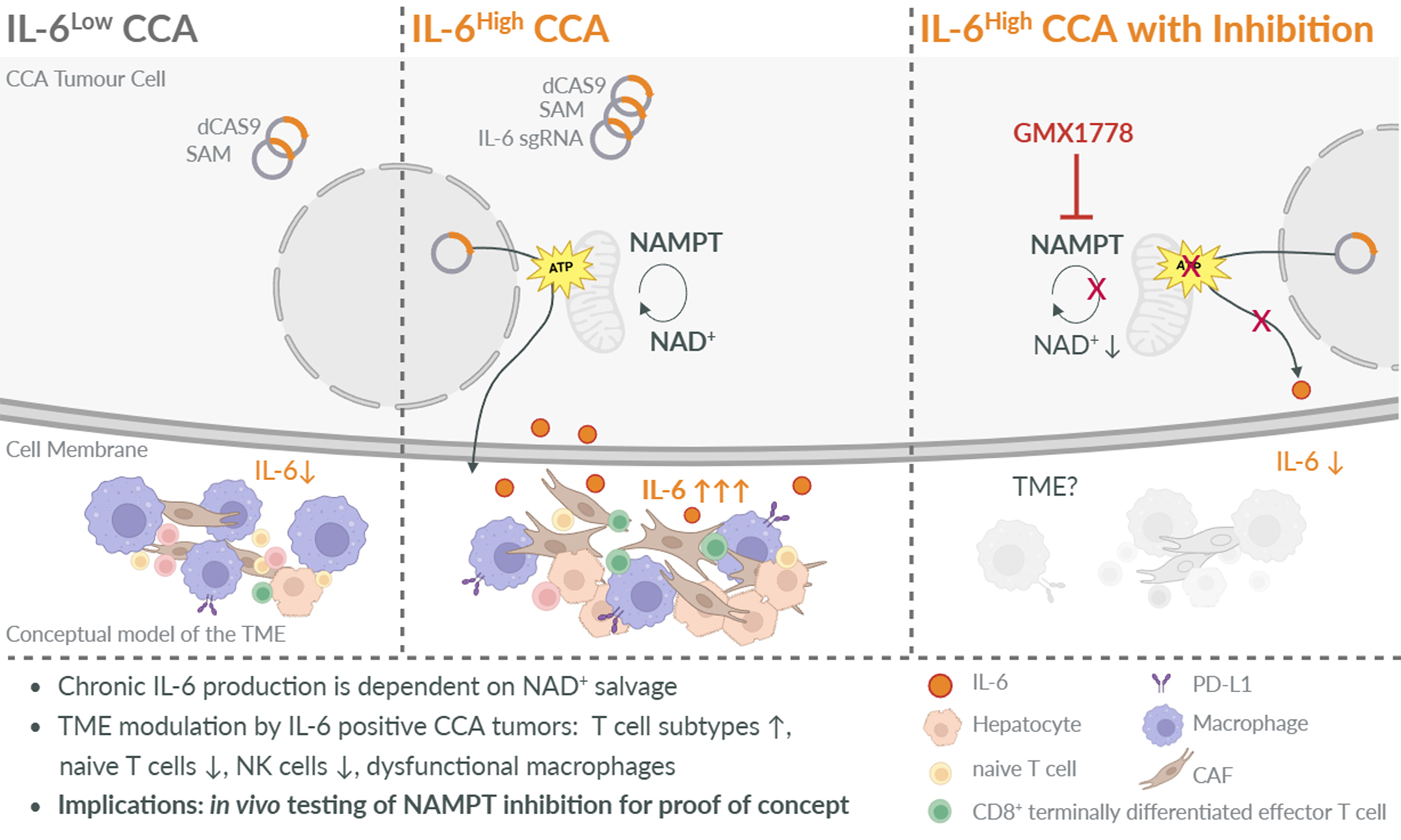
Further, the IL-6-rich CCA surrounding shapes the state of immune cells within the TME. Conditioned medium from tumor cells with high IL-6 expression (TCM IL-6) altered peripheral blood mononuclear cell (PBMC) development from healthy donors: on the one hand, TCM IL-6 enriched mature T cell subsets, whereas naïve subsets were depleted. On the other hand, tumor necrosis factor (TNF) production by T and NK cells was reduced, suggesting poor effector function. This contrasted with conditioned medium from control tumor cells (TCM dC)[1].
Furthermore, Gehl et al. observed a switch in the myeloid effector mechanism, showing that the TCM IL-6 increased the percentage of monocytes expressing IL-1β as a tumor-promoting factor-suggesting inflammasome activation[1]. Cell surface effector marker expression (CD80, CD86, CD206, CD163, MerTK) was decreased. In line with this, immunosuppressive PD-L1 was upregulated on myeloid cells. This observation supports the concept of an overall immunosuppressive switch upon tumor-cell-derived high-level IL-6 exposure[1].
Although CCA is a highly heterogeneous tumor, genetically engineered, high IL-6 signaling triggered a common drug response: three tested patient-derived cell lines showed a consistent response in a broad drug sensitivity screen. They were most sensitive to the agent GMX1778, which inhibits nicotinamide phosphoribosyltransferase (NAMPT), the rate-limiting enzyme biosynthesizing nicotinamide adenine dinucleotide (NAD+) from nicotinamide (a form of vitamin B3). NAMPT is responsible for the salvage of NAD+, the central redox partner for the main metabolic pathways (i) glycolysis; (ii) citric acid cycle; and (iii) oxidative phosphorylation. This common weak spot is not surprising, as tumor cells undergo metabolic reprogramming to meet the metabolic demands of tumor growth. Many tumor cells rely on NAMPT as a source of NAD+, as de novo synthesis from tryptophan and the Preiss-Handler pathway are more complex, and tumor cells potentially lose concerted expression of all enzymes needed for those alternative pathways upon de-differentiation[6]. At least the IL-6High tumor cells seem to depend on the salvage pathway: GMX1778 was shown to impair respiratory oxidation and reduce NAD+ levels, adenosine triphosphate (ATP) levels, and finally IL-6 expression itself[1].
The clinical landscape for IL-6 modulators is currently less clear. For CCA, one anti-IL-6 clinical trial was prematurely closed, but it did not show superiority in overall survival after 12 months with Gemcitabine/Cisplatin and Tocilizumab compared with Gemcitabine/Cisplatin alone. Potential explanations include the exclusion of an immunotherapy component rather than a quadruple combination and the lack of stratification at enrollment by IL-6 status (high/positive vs. negative). To date, no anti-IL-6 trial for intrahepatic CCA (iCCA) is registered in combination with chemo-immunotherapy, but for non-small cell lung cancer, a Phase Ib-II trial with atezolizumab (anti-PD-L1) and tocilizumab is ongoing (NCT04691817), with pending results.
NAMPT inhibition might be even more promising as it targets cancer cells’ metabolism and IL-6 production. Different inhibitors are available and have undergone early-phase clinical evaluation in other cancer types (lymphoma and melanoma). However, no therapeutic efficacy but high toxicity was documented[7], as NAD+ utilization is of general importance for any tissue and cell type. Even evaluation of a small molecule acting as a dual modulator of p21 protein (Cdc42/Rac)-activated kinase 4 (PAK4) and NAMPT for advanced solid cancers failed: combination with Niacin as rescue or Nivolumab as an immune therapy combination could not achieve an efficient anti-cancer response. Consequently, the recent trial NCT02702492 was terminated.
Potential resistance mechanisms in tumor cells could include NAD+ or NMN uptake from the extracellular space (provided by dying cells) or de novo synthesis. Of particular relevance for further in vivo testing and potential clinical implications is circadian regulation. The NAMPT gene and NAD+ levels are controlled by CLOCK, BMAL1, and SIRT1-key circadian rhythm regulators[8,9]. Applying NAMPT inhibition during the systemic physiologic nadir would allow healthy cells to sustain NAD+ levels at their peak time, whereas inhibition at the systemic nadir might still target tumor cells with their increased NAD+ need[10].
The metabolic dependency of chronic IL-6 signaling in iCCA remains to be demonstrated for clinical relevance in vivo, particularly in combination with immune therapy. As NAMPT inhibition targets the metabolic core of a cell, the addition of Gemcitabine/Cisplatin might be too toxic in humans. At least, clinical trials of NAMPT monotherapies (e.g., GMX1778 or APO866) failed to reduce tumor growth[7]. Instead, combinatorial therapies with chemotherapy (NCT00724841) led to early termination, potentially for toxicity reasons.
In addition, another aspect warrants attention: the IL-6High CCA model showed clear downregulation of MYC target genes, but accelerated activity of the MYC activation factor X (MAX). Still, MYC-targeting therapies reached significance in the drug sensitivity screen in IL-6High CCA cells[1]. The role of MAX in cancer is controversial. MAX can act as a tumor suppressor. Either as a homodimer or as a heterodimer with MD1-2, MGA, or MNT. As a partner in a heterodimer with MYC, it is tumor-promoting[11]. Hence, the downregulation of MYC, alongside increased MAX activity, is worth exploring in detail. Surprisingly, multiple distinct MYC inhibitors were effective in the drug sensitivity screen despite low MYC expression in IL-6High CCA cells[1]. As MYC and MAX are also positive regulators of the NAMPT enhancer, MYC’s downregulation but positivity in the drug sensitivity screen might point to a negative feedback loop by active NAMPT or IL-6.
In conclusion, Gehl et al. elegantly showed that induced chronic IL-6 expression in CCA tumor cells leads to their metabolic reprogramming[1]. Further, chronic IL-6 secretion by tumor cells attracts CAFs and promotes a desmoplastic and immune-suppressing TME. Artificially increased IL-6 production in CCA cells occurs at the expense of ATP and can be successfully inhibited by blocking the NAD+ salvage pathway. The intertwining of chronic, high IL-6 signaling with tumor cell metabolism suggests NAMPT inhibition as a potential additional therapeutic approach to explore in more detail in vivo. It remains unclear what role NAMPT inhibition can play across the heterogeneous landscape of CCAs and whether it may be useful for all distinct CCA types, or probably only subsets in a personalized approach, such as IL-6-positive tumors.
DECLARATIONS
Acknowledgments
The Graphical Abstract was created with BioRender.com [Created in BioRender. Heikenwälder, M. (2026) https://BioRender.com/3475gww].
Authors’ contributions
Wrote the original draft of the commentary: Hahn M
Contributed to revising the manuscript: Hahn M, Tykwe R, Heikenwälder M
Prepared the graphical abstract: Hahn M, Tykwe R
Revised the GA: Heikenwälder M
Availability of data and materials
Not applicable.
AI and AI-assisted tools statement
Not applicable.
Financial support and sponsorship
None.
Conflicts of interest
All authors declared that there are no conflicts of interest.
Ethical approval and consent to participate
Not applicable.
Consent for publication
Not applicable.
Copyright
© The Author(s) 2026.
REFERENCES
1. Gehl V, O'Rourke CJ, Cornillet M, et al. Molecular and cellular consequences of tumour-autonomous IL-6 signalling in intrahepatic cholangiocarcinoma. Gut. 2026;Epub ahead of print.
2. Banales JM, Rodrigues PM, Affò S, et al. ; International CCA Consensus Consortium. Cholangiocarcinoma 2026: status quo, unmet needs and priorities. Nat Rev Gastroenterol Hepatol. 2026;23:65-96.
3. European Association for the Study of the Liver. EASL Clinical Practice Guidelines on the management of extrahepatic cholangiocarcinoma. J Hepatol. 2025;83:211-38.
4. Høgdall D, O’Rourke CJ, Dehlendorff C, et al. Serum IL6 as a prognostic biomarker and IL6R as a therapeutic target in biliary tract cancers. Clin Cancer Res. 2020;26:5655-67.
5. Zhang M, Yang H, Wan L, et al. Single-cell transcriptomic architecture and intercellular crosstalk of human intrahepatic cholangiocarcinoma. J Hepatol. 2020;73:1118-30.
6. Garten A, Schuster S, Penke M, Gorski T, de Giorgis T, Kiess W. Physiological and pathophysiological roles of NAMPT and NAD metabolism. Nat Rev Endocrinol. 2015;11:535-46.
7. Wei Y, Xiang H, Zhang W. Review of various NAMPT inhibitors for the treatment of cancer. Front Pharmacol. 2022;13:970553.
8. Ramsey KM, Yoshino J, Brace CS, et al. Circadian clock feedback cycle through NAMPT-mediated NAD+ biosynthesis. Science. 2009;324:651-4.
9. Nakahata Y, Sahar S, Astarita G, Kaluzova M, Sassone-Corsi P. Circadian control of the NAD+ salvage pathway by CLOCK-SIRT1. Science. 2009;324:654-7.
10. Dang CV. A time for MYC: metabolism and therapy. Cold Spring Harb Symp Quant Biol. 2016;81:79-83.
Cite This Article
 Open Access
How to Cite
Download Citation
If you have the appropriate software installed, you can download article citation data to the citation manager of your choice. Simply select your manager software from the list below and click on download.
Export Citation File
Type of Import
Tips on Downloading Citation
Citation Manager File Format
Type of Import
Direct Import: When the Direct Import option is selected (the default state), a dialogue box will give you the option to Save or Open the downloaded citation data. Choosing Open will either launch your citation manager or give you a choice of applications with which to use the metadata. The Save option saves the file locally for later use.
Indirect Import: When the Indirect Import option is selected, the metadata is displayed and may be copied and pasted as needed.
About This Article
Copyright

Data & Comments
Data
0










Comments
Comments must be written in English. Spam, offensive content, impersonation, and private information will not be permitted. If any comment is reported and identified as inappropriate content by OAE staff, the comment will be removed without notice. If you have any queries or need any help, please contact us at support@oaepublish.com.