



Periareolar approach in minimally invasive thoracic surgery: a narrative review
 0
0 Abstract
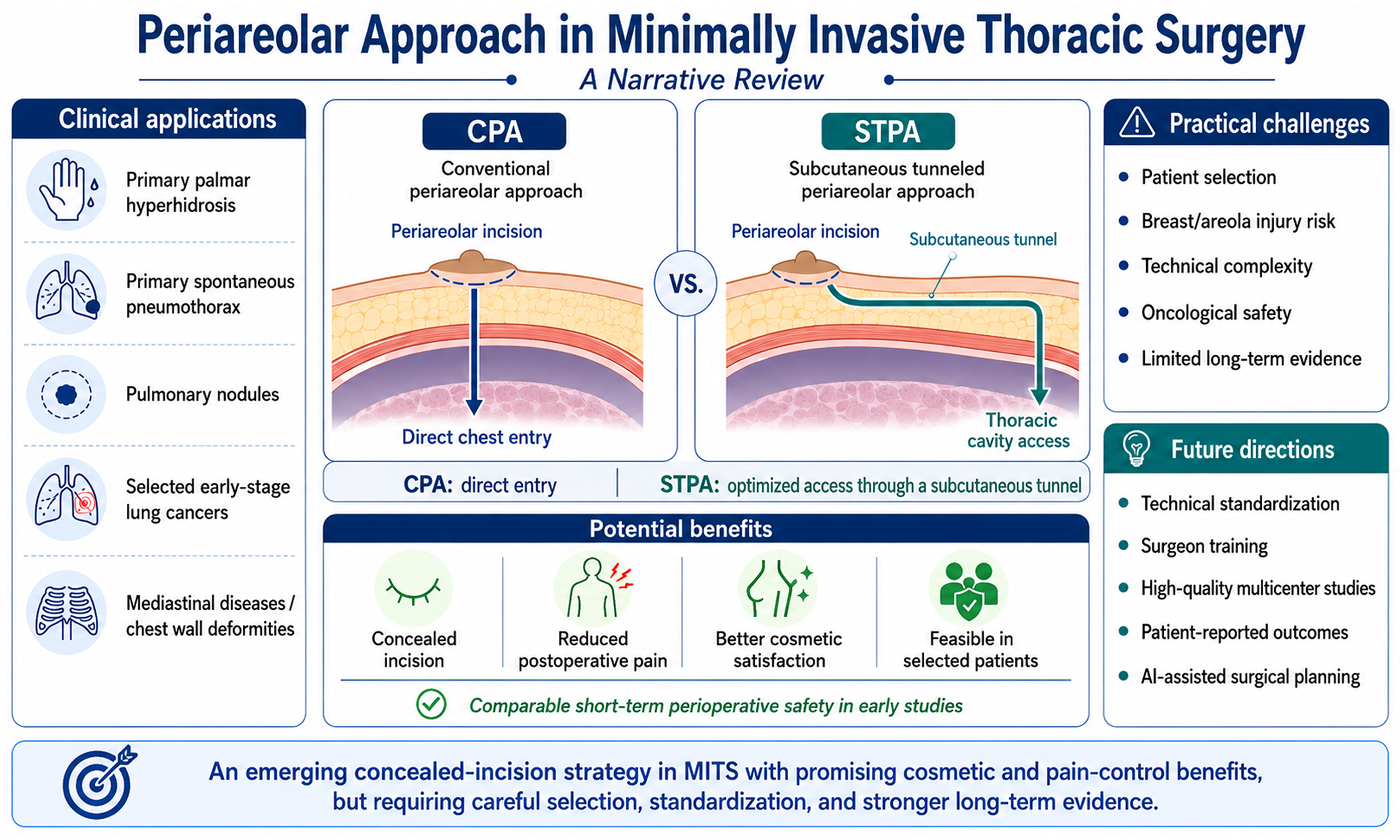
Driven by the evolving clinical demands of minimally invasive thoracic surgery, innovation in anatomical surgical approaches that promote enhanced perioperative recovery, incision concealment, and pain reduction has become an important direction for future development. As a concealed-incision strategy, the periareolar approach has been applied to a range of thoracic diseases and has shown potential advantages in cosmetic outcomes and postoperative pain control. However, broader clinical adoption remains limited by the lack of unified patient-selection criteria, insufficient standardization of surgical techniques, the risk of areolar injury, the technical learning curve, and unresolved perioperative management issues. Moreover, thoracic surgeons’ understanding of this approach remains incomplete. This review summarizes the surgical techniques, clinical applications, practical challenges, and future prospects of the periareolar approach, aiming to provide a reference for clinical practice and standardized implementation.
Keywords
INTRODUCTION
Advances in thoracoscopic techniques have driven the transition of thoracic surgery from conventional open procedures to minimally invasive approaches[1,2]. With continued improvements in surgical instruments, thoracoscopic imaging systems, and operative expertise, conventional intercostal video-assisted thoracoscopic surgery (VATS) has gradually evolved from multiport VATS to uniportal VATS[3,4]. Currently, VATS is widely used for the diagnosis and treatment of thoracic diseases involving the lung, mediastinum, and chest wall, with clear advantages in reducing surgical trauma, shortening hospital stay, and promoting postoperative recovery[5,6]. However, despite marked reductions in the number and length of VATS incisions, they are still typically placed on the lateral chest wall. Consequently, visible postoperative scarring, intercostal nerve irritation, chest wall pain, and chronic discomfort may adversely affect patients’ postoperative experience and long-term quality of life[6,7]. Therefore, while maintaining surgical safety and therapeutic efficacy, exploring more concealed, cosmetically favorable, and less invasive approaches has become an important direction in minimally invasive thoracic surgery (MITS).
The periareolar approach is a concealed-incision strategy that has attracted increasing attention in MITS[8]. This approach typically involves placing the incision along the areolar margin or within the periareolar region, thereby using the natural pigmentation boundary of the areola to conceal the postoperative scar and improve cosmetic outcomes[9]. This approach has also been adopted and refined in thyroid, breast, and cardiac surgery[10-12]. In MITS, Kesler et al. reported the use of periareolar thoracoscopic sympathectomy (TS) for primary palmar hyperhidrosis (PPH) as early as 2000, introducing this approach into thoracoscopic practice and demonstrating its potential to reduce visible chest wall scarring and improve postoperative cosmetic outcomes[13]. Since then, the periareolar approach has been applied to a range of thoracic diseases and surgical scenarios, including primary spontaneous pneumothorax (PSP), pulmonary nodules, anatomic resection for early-stage lung cancer, management of mediastinal lesions, and correction of chest wall deformities[8,14-17].
Existing evidence suggests that, in carefully selected patients with appropriate anatomical conditions, limited disease complexity, and clear expectations regarding cosmetic outcomes, the periareolar approach is technically feasible and safe and may provide better postoperative pain control and cosmetic satisfaction than conventional lateral chest wall incisions[9,18-21]. However, its application in thoracic surgery remains at an early stage, and the associated surgical techniques have not been fully standardized. Substantial heterogeneity remains across studies regarding incision design, port placement, breast tissue protection, and indication selection. Accordingly, this review summarizes the surgical techniques, clinical applications, practical challenges, and future prospects of the periareolar approach in MITS to inform rational clinical application and guide future research.
SURGICAL TECHNIQUES
Concept and patient selection
Compared with conventional lateral chest wall incisions, the periareolar approach allows a more concealed skin incision and optimized chest wall entry, thereby improving postoperative cosmetic outcomes without compromising the safety of intrathoracic manipulation[8,13,14,22].
From a technical perspective, the periareolar approach can be broadly classified into two types: the conventional periareolar approach (CPA) and the subcutaneous tunneled periareolar approach (STPA). In the CPA, the target intercostal space (ICS) is reached directly through a periareolar or circumareolar incision along the subcutaneous soft-tissue plane. In the STPA, the skin incision is placed at the areolar margin and connected to an optimal chest wall entry site through a subcutaneous tunnel. The CPA provides a shorter and more direct operative route. Therefore, it is more suitable for male patients or female patients with limited breast tissue and is commonly used for relatively simple thoracoscopic procedures, such as thoracoscopic pulmonary bullectomy (TPB) and TS[8,22-24]. In contrast, the STPA may reduce traction-related injury to the breast parenchyma, making it particularly suitable for female patients. Moreover, because the chest wall entry site can be positioned close to, or designed to replicate, a conventional thoracoscopic port site, this approach is better suited for complex procedures that require more favorable operative angles[9,20].
Appropriate patient selection is essential for the safe implementation of the periareolar approach. Therefore, patient selection should consider anatomical feasibility, disease characteristics, expected operative complexity, and patient preference, rather than cosmetic considerations alone. Current evidence suggests that STPA may be used in both male and female patients, with no clearly defined age limit[9,21,25]. According to the Chinese expert consensus, patients with good breast mobility, particularly those in whom the lateral areolar margin can be displaced toward the anterior axillary line, are considered to have favorable anatomical conditions for this approach[26]. However, the periareolar approach should be used with caution in patients with a history of ipsilateral breast or chest wall surgery, active infection at the operative site, breast disease requiring concomitant surgical management, pregnancy or lactation, suspected extensive pleural adhesions, large solid tumors, or anticipated technically complex procedures. The temporal evolution of research on the periareolar approach in MITS and the cumulative number of reported cases are illustrated in Figure 1.
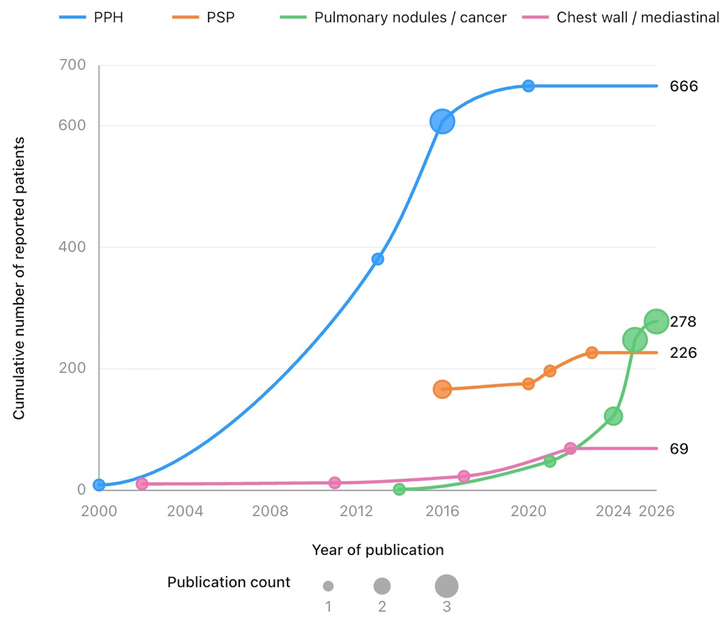
Figure 1. Research publications and cumulative reported cases of the periareolar approach in MITS (Studies with unreported sample sizes were excluded in the calculation of cumulative reported cases). Created using the Home for Researchers platform (https://www.home-for-researchers.com). MITS: Minimally invasive thoracic surgery; PPH: primary palmar hyperhidrosis; PSP: primary spontaneous pneumothorax.
Surgical procedure
The periareolar approach is typically performed under general anesthesia with double-lumen endotracheal intubation and single-lung ventilation. The patient is usually placed in the lateral decubitus position with the operative side up. Preoperative surface marking should be performed based on the locations of the lesion, areola, target ICS, auxiliary port, and chest tube exit site to define the spatial relationship between the skin incision and the chest wall entry site.
In the CPA, the incision site should be individualized based on lesion location and target ICS. In the periareolar TPB reported by Igai et al., the surgeon adjusted the patients’ upper-limb position to align the areola with the 4th ICS[27]. An approximately 2-cm incision was then made at the superior areolar margin to facilitate insertion and manipulation of the endoscopic stapler. Lin et al. selected the incision site according to the pulmonary lobe containing the nodule[24]. For nodules in the upper or middle lobe, the incision was made at the superior areolar margin corresponding to the 3rd ICS. For lower-lobe nodules, the procedure was performed through an incision at the inferior areolar margin corresponding to the 4th or 5th ICS. Therefore, the key technical principle of the CPA is to align the areolar incision as closely as possible with the target ICS through preoperative localization and patient positioning. This strategy shortens the operative route and optimizes the angle of instrument entry.
The STPA is an important modification of the CPA. The core principle is to place the skin incision at the areolar margin and create a tunnel through the subcutaneous fat layer or an appropriate anatomical plane toward the target ICS. This allows the actual chest wall entry site to be selected flexibly according to surgical requirements. Tian et al. noted that the areolar skin is relatively firm and dense[9]. Therefore, precise control of cutting force is essential to allow the incision to reach the subcutaneous fat layer directly, while avoiding repeated and unnecessary layered tissue dissection. The incision length is usually 1/3 to 1/2 of the areolar circumference. During the operation, a retractor may be used to elevate the lateral edge of the incision and provide adequate exposure of the operative field. Meticulous dissection is then performed with electrocautery along the fat plane between the lateral border of the breast tissue and the subcutaneous tissue. The dissection is extended to the region between the anterior and middle axillary lines to establish a subcutaneous tunnel toward the lateral chest wall. The thoracic cavity is usually entered through the 4th or 5th ICS. With this approach, thoracoscopic visualization and instrument manipulation can be performed through the same uniportal incision or through additional ports, depending on operative requirements. At the end of the procedure, the incision is closed in layers with 4-0 or 5-0 absorbable sutures, and the skin is closed using a cosmetic intradermal suture technique.
After completion of the intrathoracic procedure, drainage management should follow similar principles for both the CPA and STPA and should be individualized according to intraoperative air leakage and cosmetic requirements. A single 20-Fr chest tube is placed through the working port, with a preplaced suture left in situ. A water-seal test is then performed to assess pulmonary air leakage and evacuation of residual intrathoracic air. If no air leak or only minimal leakage is observed during the water-seal test, an ultrathin drainage catheter, typically an 8-Fr or 10-Fr pigtail catheter, may be used. After complete evacuation of residual intrathoracic air, the drainage catheter can be removed, and the preplaced suture can be tightened to close the incision. Use of an ultrathin drainage catheter appears to be safe and effective and may reduce postoperative pain and incision-related complications. To optimize cosmetic outcomes, the drainage catheter may be positioned away from the periareolar incision[26,28].
Management of intraoperative emergencies
In the event of an intraoperative emergency, such as massive hemorrhage caused by arterial or venous injury, prompt enlargement of the surgical access or conversion to thoracotomy may be required[9,20,28]. While the bleeding site is compressed, an additional working port can be rapidly created on the lateral chest wall, or a thoracotomy incision can be made along the 4th or 5th ICS in the axillary region. Depending on the bleeding site, the incision may be extended along the ICS to create a posterolateral or anterior thoracotomy, without extension to the periareolar incision. A rib spreader may be used to widen the ICS and provide adequate exposure of the operative field. Depending on the clinical scenario, the lung, hilum, or mediastinal structures can be exposed to facilitate vascular repair, lobectomy, or other necessary procedures.
CLINICAL APPLICATIONS
PPH
PPH was among the earliest indications for the periareolar approach in MITS [Table 1]. In 2000, Kesler et al. reported TS for severe PPH using combined axillary and periareolar incisions[13]. This technique preserved the basic principles of thoracoscopic management of the upper thoracic sympathetic chain but relocated the anterior chest wall working port to the periareolar region, thereby concealing the incision along the areolar–cutaneous junction. A subcutaneous tunnel was created from the periareolar incision to the 2nd ICS, allowing exposure, dissection, and resection of the upper thoracic sympathetic chain under thoracoscopic guidance. Lima et al. later modified this approach by performing TS through the CPA combined with a small axillary incision[29]. Compared with the earlier long periareolar incision and oblique subcutaneous tunnel, this technique reduced the incision length to 5-10 mm and limited subcutaneous dissection. Follow-up showed progressive scar fading, with no loss of nipple–areola complex sensation, infection, necrosis, or keloid formation. Pleural adhesions, large breast volume, and lactation increased technical difficulty, whereas breast implants were considered a contraindication to this approach.
Summary of studies using the periareolar approach for PPH
| Author, year | Country | Study design | Number/gender of patients | Disease/surgical procedure | Conversion rate* (%) | Subcutaneous tunnel | Incisional complications |
| Kesler et al., 2000[13] | USA | Surgical technique report | 6F 2M | PPH/TS | 12.5 (1/8) | Yes | No |
| Lima et al., 2013[29] | Brazil | Retrospective, single-arm | 372/both sexes (distribution NR) | PPH/TS | 0 | No | Hypertrophic scar: 1/372 |
| Chen et al., 2016[16] | China | Retrospective, single-arm | 85M | PPH/needlescopic thoracic sympathicotomy | 0 | No | Areolar pain: 33/85 |
| Chen et al., 2016[22] | China | Retrospective, single-arm | 58M | PPH/ETS | 0 | No | Areolar pain: 17/58 |
| Chen et al., 2016[30] | China | Randomized controlled trial (vs. conventional uniport) | 168M (periareolar: 85, control: 83) | PPH/ETS | 0 | No | Areolar pain: 3/85 |
| Lin et al., 2020[31] | China | Prospective randomized controlled trial (vs. conventional uniport) | 118M (periareolar: 58, control: 60) | PPH/ETS | 0 | No | Pneumothorax: 3/58 |
With advances in needlescopic instruments, uniportal thoracoscopy, and nonintubated anesthesia, the periareolar approach for PPH evolved from a two-port cosmetically oriented auxiliary access to a smaller-incision uniportal technique. Chen et al. reported nonintubated periareolar uniportal needlescopic thoracic sympathicotomy in 85 male patients with PPH[16]. The procedure was performed through an approximately 5-mm incision along the areolar margin using a 2-mm needle thoracoscope and an electrocautery hook. Most patients were discharged on postoperative day 1, and follow-up revealed no obvious chest wall scarring, persistent pain, or symptom recurrence. In a subsequent randomized controlled trial[30], this technique was compared with conventional transaxillary uniportal endoscopic thoracic sympathectomy (ETS). Both groups achieved immediate palmar dryness and symptom relief; however, the periareolar ETS group had a shorter incision, lower pain scores, higher cosmetic scores, and less intubation-related discomfort. Flexible endoscopy was later introduced into periareolar ETS, allowing bilateral sympathetic chain interruption through bilateral areolar-margin incision under nonintubated intravenous anesthesia. The procedure was successfully completed, with substantial improvement in PPH symptoms[22]. A 2020 prospective randomized trial[31] further showed that periareolar uniportal flexible ETS resulted in shorter incisions, lower pain scores, and higher cosmetic scores than conventional ETS, with comparable outcomes in symptom resolution, compensatory hyperhidrosis, overall satisfaction, and recurrence. Notably, except for the study by Kesler et al., which used the STPA and included 6 female patients, most early studies using the CPA involved male patients[13].
PSP
The periareolar approach was also adopted early for the surgical treatment of PSP [Table 2]. In 2016, Xu et al. reported periareolar uniportal VATS wedge resection in male patients; of the 65 patients in the periareolar group, 54 had PSP[23]. Compared with conventional VATS, the periareolar group showed no significant differences in operative time, blood loss, drainage duration, or hospital stay. However, the periareolar group had lower pain scores before and after chest tube removal, less postoperative incision-related paresthesia, and greater satisfaction with the incision. Subsequently, Lin et al. applied this approach specifically to PSP and reported periareolar TPB in 112 male patients[17]. Using a 5-mm thoracoscope, this technique involved creating a camera port and a main working port along the areolar margin for wedge resection of pulmonary bullae, air-leak testing, and postoperative drainage management. All procedures were completed successfully, with a mean operative time of 26.5 min. Most patients were discharged on postoperative days 2-3. During follow-up, no obvious chest wall scarring, persistent postoperative pain, or recurrence was reported. In 2020, Yazawa et al. conducted a prospective single-arm study in 10 male patients with PSP, further supporting the feasibility of periareolar TPB[18].
Summary of studies using the periareolar approach for PSP
| Author, year | Country | Study design | Number/gender of patients | Disease/surgical procedure | Conversion rate* (%) | Subcutaneous tunnel | Incisional complications |
| Xu et al., 2016[23] | China | Retrospective, comparative (vs. conventional two-port) | 197M (periareolar: 65, control: 132) | PSP (54/65), pulmonary nodules and bullae (11/65)/wedge resection | 0 | No | Areolar paresthesia: 39/65 |
| Lin et al., 2016[17] | China | Retrospective, single-arm | 112M | PSP, pulmonary bullae/pulmonary bullectomy | 0 | No | NR |
| Yazawa et al., 2020[18] | Japan | Prospective, single-arm clinical trial | 10M | PSP/pulmonary bullectomy | 0 | Yes (only for chest drainage) | No |
| Igai et al., 2021[27] | Japan | Prospective non-randomized, comparative (vs. conventional uniport) | 40M (periareolar: 21, control: 19) | PSP/pulmonary bullectomy | 0 | Yes (only for chest drainage) | No |
| Fu et al., 2023[19] | China | Retrospective, comparative (vs. conventional uniport) | 50M (periareolar: 29, control: 21) | PSP/pulmonary bullectomy | 0 | No | NR |
Thereafter, Igai et al. conducted a prospective comparative study in male patients with PSP, comparing the periareolar TPB (n = 21) with conventional TPB (n = 19)[27]. Perioperative outcomes and postoperative pain scores were comparable between groups. However, cosmetic satisfaction was higher in the periareolar group on postoperative day 7 (periareolar, 3.5 ± 0.6 vs. control, 2.9 ± 0.9; P = 0.02) and at postoperative month 12 (periareolar, 3.8 ± 0.5 vs. control, 3.3 ± 0.9; P = 0.0065). With advances in anesthetic techniques, tubeless strategies have been increasingly applied in MITS. In 2023, Fu et al. combined periareolar TPB with the tubeless strategy[19]. In this technique, TPB and air-leak testing were performed through an approximately 2-cm curved incision along the inferior areolar margin. A drainage tube was briefly inserted intraoperatively for negative-pressure suction and then removed immediately, after which the preplaced suture was tightened to close the incision. Compared with the conventional approach, the periareolar approach was associated with less intraoperative blood loss and lower postoperative pain scores, and no recurrence was observed in either group within 1 year. Current evidence is derived mainly from single-arm studies and small comparative cohorts, and the results should be interpreted with caution. Overall, available data suggest that short-term perioperative outcomes of the periareolar approach are broadly comparable to those of the conventional approach, with potential advantages mainly in incision concealment and cosmetic satisfaction.
Notably, the PSP-related studies summarized in this review used the CPA rather than the STPA and enrolled only male patients. Therefore, current evidence is mainly applicable to male patients with PSP who have localized lesions, no obvious pleural adhesions, and bullae suitable for periareolar manipulation.
Pulmonary nodules and early-stage lung cancers
Pulmonary nodules and early-stage lung cancer are emerging indications for the periareolar approach in MITS [Table 3]. The widespread adoption of low-dose computed tomography screening has increased the detection of early-stage lung cancers and radiologically suspicious pulmonary nodules, many of which require surgical management[32]. At the same time, patient concerns about postoperative pain, visible scarring, and quality of life have become increasingly important[33,34].
Summary of studies using the periareolar approach for pulmonary nodules and lung cancer
| Author, year | Country | Study design | Number/gender of patients | Disease/surgical procedure | Conversion rate* (%) | Subcutaneous tunnel | Incisional complications |
| Cerezo Madueño et al., 2014[8] | Spain | Case report | 1M | Pulmonary adenocarcinoma/lobectomy | 0 | No | No |
| Lin et al., 2021[24] | China | Retrospective, single-arm | 46M | Pulmonary nodules/wedge resection | 0 | No | Imperfect wound healing: 1/46 |
| Liu et al., 2024[25] | China | Retrospective, single-arm | 35F | Pulmonary nodules/wedge resection or segmentectomy | 0 | Yes | Delayed healing: 2/35 Areolar numbness: 1/35 |
| Mao et al., 2024[20] | China | Randomized, single-blind, two-arm clinical trial (vs. conventional uniport) | 29F 51M (periareolar: 40, control: 40) | Pulmonary nodules/lobectomy | 0 | Yes (only for females) | Delayed healing: 2/40 Numbness: 2/40 Mammary gland injury: 2/40 |
| Tian et al., 2025[9] | China | Surgical technique report | 4F 1M | Pulmonary nodules/wedge resection, lobectomy or segmentectomy | 0 | Yes | No |
| Yang et al., 2025[21] | China | Prospective, randomized, controlled trial (vs. conventional uniport) | 111F 63M (periareolar: 87, control: 87) | Pulmonary nodules/wedge resection, lobectomy or segmentectomy | 0 | Yes | Subcutaneous effusion: 4/87 Subcutaneous hemorrhage: 2/87 Subcutaneous stiffness: 5/87 |
| Zhang et al., 2025[35] | China | Retrospective, single-arm | 23F 10M | Lung cancer/wedge resection, lobectomy or segmentectomy | 0 | Yes | No |
| Yang et al., 2026[28] | China | Retrospective, comparative (vs. conventional uniport) | 24F 37M (periareolar: 29, control: 32) | Lung cancer/lobectomy or segmentectomy | 3.45 (1/29) | Yes | Subcutaneous hydrops: 4/29 Subcutaneous nodules: 3/29 Breast asymmetry/atrophy: 1/29 |
| Zheng et al., 2026[37] | China | Case report | 1F | Bilateral pulmonary nodules/simultaneous bilateral wedge resection | 0 | Yes | No |
| Zhang et al., 2026[36] | China | Case report | 1M | Lung cancer/bronchovascular sleeve lobectomy | 0 | Yes | No |
In 2014, Cerezo Madueño et al. first reported lobectomy using a CPA two-port VATS technique, demonstrating its technical feasibility and favorable cosmetic outcomes[8]. Subsequent studies from China in 2016 and 2021 applied this approach to patients with pulmonary nodules and further supported its safety and cosmetic advantages[23,24]. However, CPA techniques generally accessed the thoracic cavity directly through the periareolar incision, which carried a potential risk of breast tissue injury. As a result, their application was largely limited to male patients.
In 2024, Liu et al. modified the CPA by introducing a subcutaneous tunneled technique[25]. By creating a subcutaneous tunnel between the periareolar incision and the thoracic cavity, this technique avoids direct traversal of breast tissue and has been successfully applied in female patients with pulmonary ground-glass nodules. In the same year, the group conducted a single-blind randomized controlled trial in patients undergoing VATS right middle lobectomy[20]. Compared with conventional uniportal VATS, the periareolar approach achieved better postoperative pain control and cosmetic outcomes, with comparable blood loss (periareolar, 68 ± 31 mL vs. control, 84 ± 77 mL; P > 0.05) and complication rates (periareolar, 30.0% vs. control, 37.5%; P > 0.05), and no cases requiring conversion to thoracotomy.
The emergence and refinement of the subcutaneous tunneled technique have further expanded the indications for the periareolar approach. In 2025, Tian et al. systematically described the surgical workflow of STPA uniportal VATS pulmonary resection, based on prior experience with intercostal VATS and CPA pulmonary resection[9]. Their work provided a practical reference for standardizing and disseminating the technique and further supported its application across different types of pulmonary resection and in patients of both sexes. Subsequently, the first prospective randomized controlled study of the STPA involved 174 patients undergoing pulmonary resection[21]. The study showed that this approach had perioperative safety comparable to conventional uniportal VATS, while offering clear advantages in scar assessment and cosmetic satisfaction. Similar findings were subsequently reported in several retrospective cohort studies[28,35]. Notably, in the study by Yang et al., one patient with severe pleural adhesions after neoadjuvant therapy required conversion to thoracotomy, suggesting that this approach remains compatible with standard thoracic surgical principles for conversion in complex cases[28].
With accumulating clinical experience, the STPA has gradually expanded from simple pulmonary resections to more complex thoracic procedures. Recent case reports have described the successful use of this technique for non-intubated simultaneous bilateral pulmonary nodule resection and bronchovascular sleeve lobectomy for centrally located lung squamous cell carcinoma[36,37]. Although current evidence remains limited to case reports, these exploratory applications suggest the technical feasibility and potential value of the STPA for complex pulmonary resections.
Overall, the available evidence provides preliminary support for the safety and feasibility of the periareolar approach in the surgical management of pulmonary nodules and selected early-stage lung cancers, with potential benefits in postoperative pain control and cosmetic outcomes. Although early results are encouraging, current evidence is derived predominantly from single-center studies, particularly in China, and appropriate patient selection and long-term oncological assessment remain essential before this approach can be widely adopted for malignant thoracic diseases.
Chest wall deformities and mediastinal diseases
The periareolar approach has also been explored for repairing chest wall deformities [Table 4]. A previous report showed that, in patients with pectus excavatum, a modified Nuss procedure could be performed under bilateral thoracoscopic guidance through small periareolar or submammary incisions, allowing insertion of a shorter, less curved corrective bar[15]. Combined with sternal transection, this approach may reduce chest wall tension and facilitate sternal elevation, thereby improving deformity correction and cosmetic outcomes. However, this technique appears more suitable for symmetric deformities; in patients with marked asymmetry or complex chest wall rigidity, the operative approach should be individualized. Subsequently, Innocenti et al. applied the periareolar approach to custom-made silicone implant placement for aesthetic correction of pectus excavatum[38]. In their series of 11 adult patients, no major complications or implant displacement were observed, although postoperative seroma occurred in 6 patients. These findings suggest that this method is primarily an aesthetic volume-restoration procedure and may be more suitable for patients without significant cardiopulmonary impairment and with relatively localized deformities. A similar concealed-incision concept has also been applied to pectus carinatum repair, in which a modified bar was inserted through small periareolar or submammary incisions, achieving favorable scar concealment[39]. However, the sample size was small, and the available evidence remains largely limited to technical reports.
Summary of studies using the periareolar approach for chest wall deformities and mediastinal diseases
| Author, year | Country | Study design | Number/gender of patients | Disease/surgical procedure | Conversion rate* (%) | Subcutaneous tunnel | Incisional complications |
| Pérez et al., 2011[39] | Spain | Surgical technique report | 1F 1M | Pectus carinatum/modified Nuss procedure | 0 | No | No |
| Pérez et al., 2011[15] | Spain | Surgical technique report | NR | Pectus excavatum/modified Nuss procedure combined with sternotomy | NR | NR | NR |
| Innocenti et al., 2017[38] | Italy | Retrospective case series | 3F 8M | Pectus excavatum/silicone sternal implant | NR | Yes | Seroma: 6/11 |
| Tabet et al., 2002[14] | Lebanon | Surgical technique report | 10F | Anterior mediastinal or aortopulmonary nodes/left anterior mediastinotomy | 0 | Yes | Left internal mammary artery injury: 1/10 |
| Lin et al., 2022[40] | China | Retrospective, single arm | 46M | Thymic disease with myasthenia gravis/extended thymectomy | 0 | No | Areolar pain: 5/46 |
For mediastinal diseases, the periareolar approach has primarily been used for mediastinal biopsy and extended thymectomy [Table 4]. Tabet et al. previously reported a periareolar mediastinoscopic technique[14]. This approach provides access to the anterior mediastinum or aortopulmonary window through a small incision in the superomedial aspect of the left areola. Histopathological diagnoses were successfully obtained in all 10 young female patients with cosmetic concerns. In that study, 1 patient sustained an internal mammary artery injury, which was managed through the original incision; no nipple-areolar sensory disturbance or residual pneumothorax was observed. More recently, Lin et al. reported uniportal thoracoscopic extended thymectomy in 46 male patients with myasthenia gravis via the periareolar approach[40]. All procedures were completed successfully, with no conversion to thoracotomy and no operative mortality. The mean postoperative drainage volume was 63.4 ± 28.2 mL/day, and the mean drainage duration was 1.6 ± 1.2 days. Postoperative incisions were well concealed; patients experienced mild postoperative pain and some symptomatic improvement.
Overall, the periareolar approach appears to offer favorable cosmetic advantages in selected patients with chest wall deformities or mediastinal diseases. However, current evidence is largely derived from small, single-center studies with strict patient-selection criteria. Therefore, broader adoption of this approach should be considered cautiously.
PRACTICAL CHALLENGES
Risk of breast and areolar injury
One major safety concern associated with the periareolar approach is potential injury to breast tissue and the nipple-areola complex, which represents a unique consideration compared with conventional VATS approaches[9,25,27,28]. This issue is particularly relevant in female patients, because an incision trajectory that directly traverses the breast parenchyma may increase the risk of tissue injury, altered areolar sensation, local induration, and postoperative imaging changes. However, considerable heterogeneity exists among current studies regarding patient selection, as most early reports focused on male patients, whereas recent studies have expanded the application of the STPA to selected female patients with favorable breast anatomy. Therefore, when creating a subcutaneous tunnel, careful dissection should be performed along a relatively safe subcutaneous plane, with avoidance of excessive traction or compression of breast tissue. Intraoperatively, particular attention should be paid to preserving the vascular supply and sensory innervation of the nipple-areola complex. In addition to routine postoperative assessment of wound healing and bleeding, areolar sensation, local breast induration, subcutaneous fluid accumulation, and breast-related imaging findings should be monitored[26].
Moreover, because the skin at the areolar margin is relatively thin, prolonged instrument placement or repeated manipulation with multiple instruments may lead to local ischemia, skin contusion, wound dehiscence, or scar widening. During wound closure, layered suturing of the chest wall and skin should be performed, and excessive tension at the areolar margin should be avoided to reduce the risk of areolar deformity and impaired wound healing[9,25]. When operative access is established through a subcutaneous tunnel, moderate external compression of the tunnel area, for example, using a postoperative chest binder, may help reduce complications such as subcutaneous fluid accumulation and fat liquefaction[26].
Surgical trajectory and operative limitations
The skin location of a periareolar incision is primarily constrained by areolar anatomy, whereas the optimal chest wall entry point depends on lesion location, the surgical trajectory, and hilar anatomy[28]. These two requirements do not always coincide. Moreover, variations in incision design, subcutaneous tunnel creation, and port configuration among different studies may contribute to differences in technical difficulty and reported outcomes. Attempting to perform complex hilar procedures through a single periareolar incision may result in an unfavorable trocar angle, instrument interference, excessive vascular traction, or a restricted stapling direction, thereby increasing operative risk. For lobectomy, complex segmentectomy, or procedures requiring extensive hilar manipulation, surgeons should consider adding an auxiliary port based on hilar exposure, fissure development, vascular anatomy, and specimen size, rather than prioritizing incision concealment alone[21]. In cases involving severe pleural adhesions, unclear hilar anatomy, tumor invasion of adjacent structures, or anticipated difficulty in safely managing the vessels or bronchus, timely conversion to conventional VATS or open surgery should be considered.
Oncological safety
For benign lesions or bullectomy, the resected specimen is usually small and can often be retrieved smoothly through the periareolar incision. However, in malignant tumor surgery, specimen retrieval requires stricter control to ensure oncological safety. The specimen should be removed using an endoscopic retrieval bag to minimize the risk of wound contamination and potential tumor dissemination. If the specimen is large, forceful extraction through a small incision should not be attempted solely to preserve incision concealment. Instead, enlarge the incision when necessary, or select an alternative retrieval route.
In malignant tumor surgery, complete resection, adherence to the no-touch principle, adequate surgical margins, and systematic lymph node dissection should always take precedence over cosmetic considerations. At present, evidence on the periareolar approach for lung cancer remains limited, and long-term recurrence, survival outcomes, and oncological equivalence with conventional VATS approaches have not been sufficiently evaluated. Future studies should further clarify whether this approach can achieve comparable oncological outcomes in terms of surgical margins, lymph node dissection quality, and long-term survival. Therefore, in current clinical practice, short-term technical feasibility should not be directly equated with long-term oncological safety.
Perioperative management
Perioperative management of the periareolar approach should encompass routine thoracic surgical care as well as dynamic assessment of pain, scarring, and areola-related complications[10,21,25,41]. Adequate preoperative communication may help patients understand the potential benefits and limitations of this approach, improve treatment adherence, and support individualized surgical decision-making. Postoperatively, pain intensity can be dynamically assessed using standardized pain scales, and wound healing around the areolar incision, hypertrophic scarring, local sensory changes, and subcutaneous fluid accumulation should be closely monitored[21,28]. Pain management should follow enhanced recovery after surgery principles. A multimodal analgesic strategy, including non-opioid analgesics, local anesthetic infiltration, and intercostal nerve blockade when appropriate, may further reduce postoperative pain and facilitate early recovery. However, standardized pain management protocols specifically tailored to the periareolar approach remain lacking and warrant further investigation.
Chest tube management is an often overlooked but clinically important aspect of the periareolar approach[42]. Direct placement of the chest tube through the periareolar incision may compromise wound cosmesis and increase the risks of local areolar traction, pain, and infection[35]. Therefore, when an auxiliary port is available, chest tube placement through this port should be preferred. For periareolar uniportal VATS procedures, a small-bore chest tube, short-term drainage, or an individualized tubeless strategy may be selected based on the procedure type, risk of air leakage, pleural drainage volume, and postoperative recovery[26]. However, the safety of these strategies and the patient populations most likely to benefit from them require further investigation. Future studies should focus on establishing standardized perioperative management protocols, including pain control strategies, drainage management, and postoperative rehabilitation.
FUTURE PROSPECTS
Technical standardization and training
The periareolar approach was initially developed in the United States and subsequently adopted in Europe, primarily for male patients. After the introduction of the STPA concept in 2024, its application expanded to female patients, with rapid adoption in China. Currently, Chinese centers account for approximately 65% of reported cases. The technique remains in an early phase of clinical development, and further standardization is essential for its broader application[9]. Future studies should clarify key technical elements, including patient selection, incision design, tunnel creation, chest wall entry site, drainage strategy, specimen retrieval, and emergency conversion. In particular, the STPA learning curve requires further evaluation. Stepwise training, simulation-based education, and initial application in carefully selected simple cases may help improve procedural safety and reproducibility before extension to complex anatomical resections[21].
High-quality clinical evidence
Current evidence is mainly derived from single-center studies, retrospective cohorts, single-arm studies, small prospective series, and early randomized trials[20-23]. Although available data suggest potential advantages in scar concealment, postoperative pain control, and cosmetic satisfaction, long-term outcomes remain insufficiently evaluated. Future multicenter prospective studies and randomized controlled trials are needed to compare the periareolar approach with conventional uniportal VATS and robotic-assisted thoracic surgery. For lung cancer surgery, long-term oncological outcomes, including margin status, lymph node dissection quality, recurrence, disease-free survival, and overall survival, should be systematically assessed.
Patient-reported outcomes and individualized selection
The value of the periareolar approach should not be judged solely by perioperative safety or cosmetic appearance. Future research should incorporate standardized patient-reported outcomes, including chronic pain, scar satisfaction, body image, nipple-areola sensation, quality of life, and psychological acceptance[33,43]. This approach should not be considered a universal substitute for conventional VATS, but rather a selective option for anatomically suitable patients with clear cosmetic or quality-of-life concerns. Shared decision-making is therefore important to balance incision concealment against potential risks, operative complexity, and uncertain long-term benefits.
Artificial intelligence-assisted surgical planning
Artificial intelligence (AI) and digital surgical planning may further improve the precision and safety of the periareolar approach. Preoperative three-dimensional reconstruction, AI-assisted image segmentation, and virtual simulation may facilitate individualized planning by clarifying the spatial relationship among the pulmonary lesion, hilar structures, chest wall entry site, and planned resection margin, and may assist in optimizing the design of the periareolar incision and subcutaneous tunnel[44-47]. These tools may be particularly useful for complex segmentectomy, deep pulmonary nodules, bilateral procedures, and patients with anatomical variations. In the future, AI-based decision-support systems may also assist in patient selection by integrating lesion location, breast morphology, surgical complexity, and patient preference.
Integration with emerging minimally invasive platforms
The combination of the periareolar approach with robotic systems, flexible endoscopy, needlescopic instruments, and novel navigation technologies deserves further exploration[48]. Robotic platforms may overcome some technical limitations of the periareolar approach by providing three-dimensional visualization, enhanced dexterity, and articulated instrumentation, although dedicated clinical evaluation remains lacking[49-51]. However, these applications require careful assessment of port configuration, external instrument collision, tunnel safety, cost-effectiveness, and oncological reliability. The future development of this approach should prioritize surgical safety and reproducibility rather than cosmetic benefit alone.
CONCLUSIONS
The periareolar approach is an emerging concealed-incision strategy in MITS. Current evidence suggests that, in carefully selected patients, this approach is feasible and safe and has been applied to PPH, PSP, pulmonary nodules, and selected lung cancers. Compared with conventional thoracoscopic incisions, it may offer advantages in scar concealment, postoperative pain control, and cosmetic satisfaction without an apparent increase in perioperative complications. However, its broader application remains limited by patient selection, the risk of areolar injury, technical complexity, and insufficient long-term oncological evidence. In clinical practice, the optimal surgical strategy should be determined according to the patient’s condition and individualized needs, with surgical safety remaining the foremost principle.
DECLARATIONS
Acknowledgments
The graphical abstract was created using the Home for Researchers platform (https://www.home-for-researchers.com).
Authors’ contributions
Conceptualization: Wang J (Jia Wang), Lin P, Wang J (Junjie Wang), Tian D
Literature search and data curation: Wang J (Jia Wang), Lin P, Wang J (Junjie Wang), Tian D
Analysis and interpretation: Wang J (Jia Wang), Lin P, Wang J (Junjie Wang), Tian D
Writing - original draft: Wang J (Jia Wang), Lin P
Writing - review and editing: Wang J (Junjie Wang), Tian D
Supervision: Zhou H, Chen L
Funding acquisition: Tian D
All authors reviewed and approved the final version of the manuscript.
Availability of data and materials
Not applicable.
AI and AI-assisted tools statement
Not applicable.
Financial support and sponsorship
None.
Conflicts of interest
All authors declare that there are no conflicts of interest.
Ethical approval and consent to participate
Not applicable.
Consent for publication
Not applicable.
Copyright
© The Author(s) 2026.
REFERENCES
1. Kirby TJ, Mack MJ, Landreneau RJ, Rice TW. Lobectomy - video-assisted thoracic surgery versus muscle-sparing thoracotomy. A randomized trial. J Thorac Cardiovasc Surg. 1995;109:997-1001; discussion 1001.
2. Flores RM, Park BJ, Dycoco J, et al. Lobectomy by video-assisted thoracic surgery (VATS) versus thoracotomy for lung cancer. J Thorac Cardiovasc Surg. 2009;138:11-8.
3. Rocco G, Martin-Ucar A, Passera E. Uniportal VATS wedge pulmonary resections. Ann Thorac Surg. 2004;77:726-8.
4. Gonzalez D, Paradela M, Garcia J, Dela Torre M. Single-port video-assisted thoracoscopic lobectomy. Interact Cardiovasc Thorac Surg. 2011;12:514-5.
5. Wildgaard K, Ravn J, Kehlet H. Chronic post-thoracotomy pain: a critical review of pathogenic mechanisms and strategies for prevention. Eur J Cardiothorac Surg. 2009;36:170-80.
6. Bendixen M, Jørgensen OD, Kronborg C, Andersen C, Licht PB. Postoperative pain and quality of life after lobectomy via video-assisted thoracoscopic surgery or anterolateral thoracotomy for early stage lung cancer: a randomised controlled trial. Lancet Oncol. 2016;17:836-44.
7. Qsous G, Downes A, Carroll B, et al. A comparison of the differences in postoperative chronic pain between video-assisted and robotic-assisted approaches in thoracic surgery. Cureus. 2022;14:e31688.
8. Cerezo Madueño F, Arango Tomás E, Algar Algar FJ, Salvatierra Velázquez A. Periareolar approach for thoracoscopic lobectomy. Ann Thorac Surg. 2014;97:1427-9.
9. Tian D, Wang JJ, Jiang KY, et al. The subcutaneous tunneled periareolar approach in lung resection performed through uniportal video-assisted thoracic surgery. J Thorac Dis. 2025;17:1073-9.
10. Klinger M, Vinci V, Giannasi S, et al. The periareolar approach: all seasons technique for multiple breast conditions. Plast Reconstr Surg Glob Open. 2021;9:e3693.
11. Van Praet KM, Kofler M, Akansel S, et al. Periareolar endoscopic minimally invasive cardiac surgery: postoperative scar assessment analysis. Interact Cardiovasc Thorac Surg. 2022;35:ivac200.
12. Choi YS, Choi JH, Jeon MS, et al. First experience of single-port robotic areolar approach thyroidectomy. Clin Exp Otorhinolaryngol. 2023;16:275-81.
13. Kesler KA, Brooks-Brunn JA, Campbell RL, Brown JW. Thoracoscopic sympathectomy for hyperhidrosis palmaris: a periareolar approach. Ann Thorac Surg. 2000;70:314-7.
14. Tabet GS, Kassabian EB, Rohayem JN, Jebara VA. Periareolar mediastinoscopy: a new technique. Ann Thorac Surg. 2002;73:1335-6.
15. Pérez D, Cano JR, Quevedo S, López L. Videothoracoscopic repair of pectus excavatum with sternal transection for adult patients with nonelastic deformity. J Thorac Cardiovasc Surg. 2011;142:942-3.
16. Chen JF, Lin JB, Tu YR, et al. Nonintubated transareolar single-port thoracic sympathicotomy with a needle scope in a series of 85 male patients. Surg Endosc. 2016;30:3447-53.
17. Lin JB, Chen JF, Lai FC, Li X, Qiu ML. Transareolar pulmonary bullectomy for primary spontaneous pneumothorax. J Thorac Cardiovasc Surg. 2016;152:999-1005.
18. Yazawa T, Igai H, Ohsawa F, Yoshikawa R, Matsuura N, Kamiyoshihara M. Feasibility of thoracoscopic pulmonary bullectomy using a transareolar approach for treatment of primary spontaneous pneumothorax. J Thorac Dis. 2020;12:5794-801.
19. Fu H, Jin D, Wei Y. Thoracoscopic treatment of primary spontaneous pneumothorax without a drainage tube in male patients. J Int Med Res. 2023;51:3000605231154394.
20. Mao Z, Dong P, Zhou Q, Zhang S. Periareolar approach in video-assisted thoracoscopic surgery for right middle lobectomy: a novel technique. Surg Endosc. 2024;38:5457-63.
21. Yang Z, Chen M, Wu J, et al. Comparison of subcutaneous tunnel-assisted periareolar incision versus conventional uniportal video-assisted thoracoscopic surgery (VATS) for pulmonary nodules: a prospective, randomized, controlled trial. Eur J Surg Oncol. 2025;51:110353.
22. Chen J, Lin J, Tu Y, Lin M, Li X, Lai F. Nonintubated transareolar endoscopic thoracic sympathectomy with a flexible endoscope: experience of 58 cases. Ann Thorac Cardiovasc Surg. 2016;22:12-9.
23. Xu K, Bian W, Xie H, Ma H, Ni B. Single-port video-assisted thoracoscopic wedge resection: novel approaches in different genders. Interact Cardiovasc Thorac Surg. 2016;23:202-7.
24. Lin J, Yin Y, Zhuo Y, Li X, Lai F. Transareolar uniportal video-assisted thoracoscopic surgery for the treatment of male patients with peripheral pulmonary nodules: a novel technique in thoracic surgery. Updates Surg. 2021;73:1541-8.
25. Liu Y, Dong P, Zhang S, Geng Q, Mao Z. Transareolar video-assisted thoracoscopic surgery in females: a novel incision for pulmonary ground glass nodule resection. J Laparoendosc Adv Surg Tech A. 2024;34:490-6.
26. Zheng B, Tian D, Mao Z. Expert consensus on periareolar concealed uniportal video-assisted thoracoscopic lung resection. Chin J Clin Thorac Cardiovasc Surg. 2025;32:731-7. (in Chinese).
27. Igai H, Kamiyoshihara M, Furusawa S, Ohsawa F, Yazawa T, Matsuura N. A prospective comparative study of thoracoscopic transareolar and uniportal approaches for young male patients with primary spontaneous pneumothorax. Gen Thorac Cardiovasc Surg. 2021;69:1414-20.
28. Yang K, Li K, Guo C, et al. Analysis of the safety and feasibility of subcutaneous areolar tunnel single-port thoracoscopic radical resection for lung cancer: a retrospective cohort study. Updates Surg. 2026.
29. Lima SO, Abuawad YG, Santos PS, Pinto Neto AF, de Santana VR, Reis FP. Infraareolar access for thoracoscopic sympathectomy to treat primary hyperhidrosis. Surg Today. 2013;43:221-4.
30. Chen J, Du Q, Lin M, et al. Transareolar single-port needlescopic thoracic sympathectomy under intravenous anesthesia without intubation: a randomized controlled trial. J Laparoendosc Adv Surg Tech A. 2016;26:958-64.
31. Lin JB, Kang MQ, Chen JF, et al. Transareolar single-port endoscopic thoracic sympathectomy with a flexible endoscope for primary palmar hyperhidrosis: a prospective randomized controlled trial. Ann Transl Med. 2020;8:1659.
32. Yotsukura M, Asamura H, Motoi N, et al. Long-term prognosis of patients with resected adenocarcinoma in situ and minimally invasive adenocarcinoma of the lung. J Thorac Oncol. 2021;16:1312-20.
33. Batchelor TJP, Rasburn NJ, Abdelnour-Berchtold E, et al. Guidelines for enhanced recovery after lung surgery: recommendations of the Enhanced Recovery After Surgery (ERAS®) Society and the European Society of Thoracic Surgeons (ESTS). Eur J Cardiothorac Surg. 2019;55:91-115.
34. Merlo A, Carlson R, Espey J 3rd, et al. Postoperative symptom burden in patients undergoing lung cancer surgery. J Pain Symptom Manage. 2022;64:254-67.
35. Zhang Z, Yu X, Peng X, Fang J. Single-port thoracoscopic surgery via areola approach for lung cancer: feasibility, safety, and initial experience with a novel technique. ANZ J Surg. 2025;95:2367-72.
36. Zhang J, Zhang Z, Zhang J, Yu X, Fang J. Uniportal transareolar video-assisted thoracoscopic surgery bronchovascular sleeve lobectomy for central squamous cell carcinoma: a case report. JTCVS Tech. 2026;38:102403.
37. Zheng J, Wang C, Zhang D, Tay Y, Li S. Simultaneous bilateral nonintubated uniportal video-assisted thoracoscopic surgery via transareolar approach for multiple pulmonary nodules. JTCVS Tech. 2026;36:102275.
38. Innocenti A, Ciancio F, Melita D, et al. Periareolar access for pectus excavatum correction with silicone implants: a new method to minimize postoperative scars-review of the literature, considerations and statistical analysis of clinical outcomes. Aesthetic Plast Surg. 2017;41:878-86.
39. Pérez D, Cano JR, Quevedo S, López L. New minimally invasive technique for correction of pectus carinatum. Eur J Cardiothorac Surg. 2011;39:271-3.
40. Lin J, Lin N, Li X, Lai F. Transareolar uniportal thoracoscopic extended thymectomy for patients with myasthenia gravis. Front Surg. 2022;9:914677.
41. Sun J, Liu C, Mu D, et al. Chinese women’s preferences and concerns regarding incision location for breast augmentation surgery: a survey of 216 patients. Aesthetic Plast Surg. 2015;39:214-26.
42. Holbek BL, Huang L, Christensen TD, et al. Efficacy of avoiding chest drains after video-assisted thoracoscopic surgery wedge resection: protocol for a randomised controlled trial. BMJ Open. 2024;14:e080573.
43. Dai W, Wang Y, Liao J, et al. Electronic patient-reported outcome-based symptom management versus usual care after lung cancer surgery: long-term results of a multicenter, randomized, controlled trial. J Clin Oncol. 2024;42:2126-31.
44. Li X, Zhang S, Luo X, et al. Accuracy and efficiency of an artificial intelligence-based pulmonary broncho-vascular three-dimensional reconstruction system supporting thoracic surgery: retrospective and prospective validation study. EBioMedicine. 2023;87:104422.
45. Chen X, Dai C, Peng M, et al. Artificial intelligence driven 3D reconstruction for enhanced lung surgery planning. Nat Commun. 2025;16:4086.
46. He H, Yu C, Yang Y, Maessen JG, Sardari Nia P. Three-dimensional reconstruction and virtual simulation of patient-specific anatomy for procedural planning in thoracoscopic segmentectomy: a systematic review and meta-analysis. Eur J Cardiothorac Surg. 2025;67:ezaf283.
47. Liang H, Yan Z, Zhang Y, et al. LungSurg: a generative AI system for segmentation and phase classification in thoracoscopic lobectomy. MedComm. 2026;7:e70613.
48. Wang W, Zhang S, Dong P, Mao Z. Initial experience with a novel 2-mm needle-type intracorporeal assembly forceps for periareolar video-assisted thoracoscopic surgery. Surg Endosc. 2025;39:7814-21.
49. Niu Z, Cao Y, Du M, et al. Robotic-assisted versus video-assisted lobectomy for resectable non-small-cell lung cancer: the RVlob randomized controlled trial. EClinicalMedicine. 2024;74:102707.
50. Li C, Chen X, Wang X, et al. Single-port robotic-assisted approach in thoracic surgery: a prospective real-world study. Interdiscip Cardiovasc Thorac Surg. 2025;40:ivaf161.
Cite This Article
 Open Access
How to Cite
Download Citation
Export Citation File:
Type of Import
Tips on Downloading Citation
Citation Manager File Format
Type of Import
Direct Import: When the Direct Import option is selected (the default state), a dialogue box will give you the option to Save or Open the downloaded citation data. Choosing Open will either launch your citation manager or give you a choice of applications with which to use the metadata. The Save option saves the file locally for later use.
Indirect Import: When the Indirect Import option is selected, the metadata is displayed and may be copied and pasted as needed.
About This Article
Copyright

Data & Comments
Data
0






Comments
Comments must be written in English. Spam, offensive content, impersonation, and private information will not be permitted. If any comment is reported and identified as inappropriate content by OAE staff, the comment will be removed without notice. If you have any queries or need any help, please contact us at support@oaepublish.com.