

Function-preserving strategies for localized prostate cancer: minimally invasive radical surgery, focal therapy and AI-assisted surgery
 0
0 
Graphical Abstract
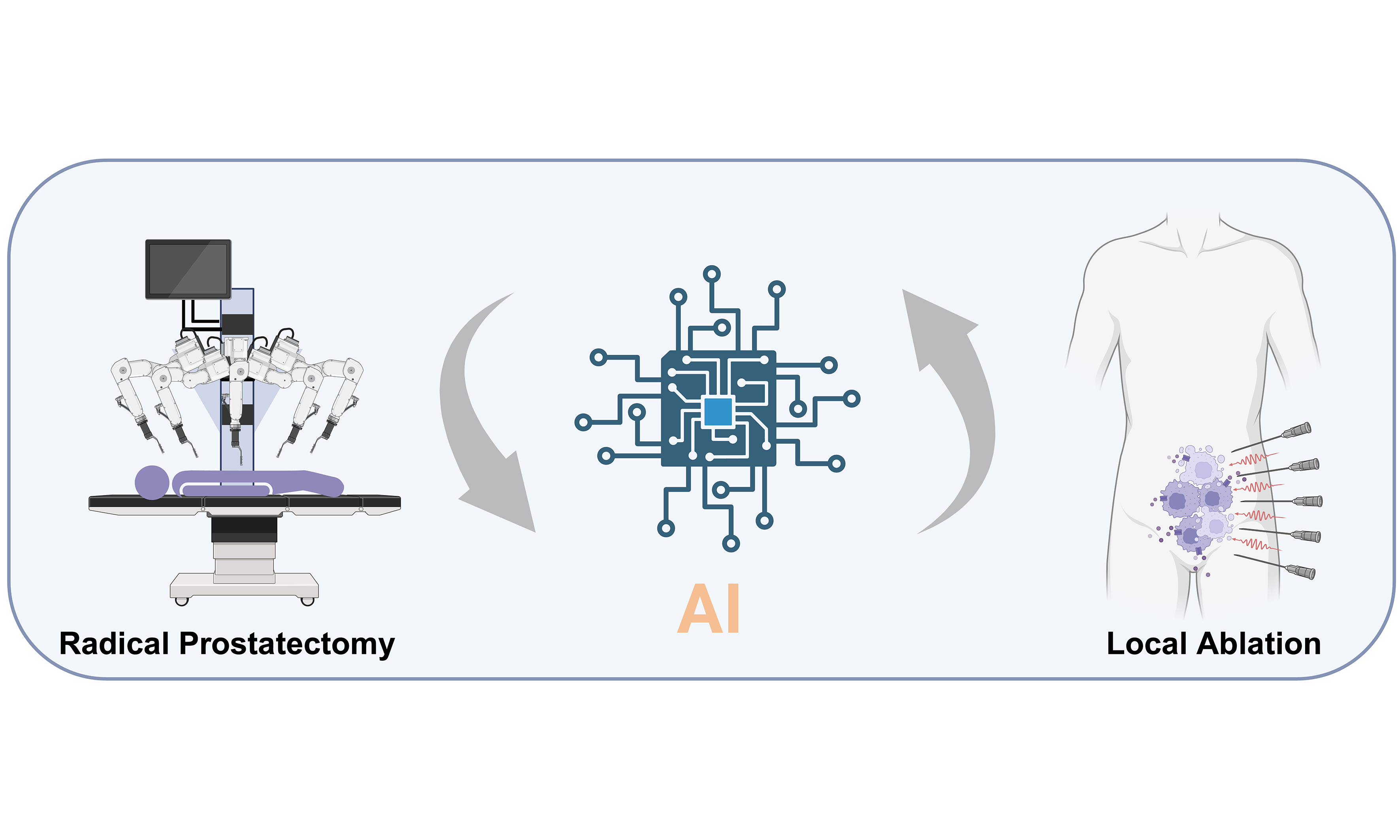
Abstract
With increasing detection and longer survival in localized prostate cancer (PCa), treatment goals increasingly emphasize preserving urinary continence and sexual function without compromising oncological control. This review synthesizes evidence on function-preserving strategies, focusing on minimally invasive radical prostatectomy, focal ablation, and artificial intelligence (AI)-assisted surgery. Laparoscopic and robot-assisted radical prostatectomy remain the principal radical approaches; multiport robot-assisted surgery has the most mature evidence for balancing cancer control and functional recovery. Relevant refinements include nerve-sparing, fluorescence imaging, three-dimensional reconstruction, and AI-assisted risk stratification, planning, navigation, and outcome prediction. In carefully selected patients with low- or intermediate-risk disease, focal therapies may reduce treatment burden and preserve function. Focal therapy is not yet a standard option for localized prostate cancer, but it may be discussed during shared decision-making with carefully selected, fully informed patients. Some focal modalities are already used in selected clinical settings, including outside formal trials where locally permitted, although implementation varies across health-care systems. Counseling should address limitations in long-term evidence, the need for structured surveillance, and possible repeat or salvage treatment. Heterogeneous selection, endpoints, and follow-up, together with a lack of high-quality head-to-head comparisons, preclude reliable ranking of focal modalities; sexual recovery and long-term oncological durability also vary. Direct evidence that AI improves nerve preservation, continence, erectile function, or margin control remains insufficient for routine adoption. Optimal function preservation requires risk-adapted selection, technical expertise, multidisciplinary decision-making, and rigorous follow-up. Future studies should prospectively validate AI tools and standardize imaging, outcomes, and clinically meaningful endpoints.
Keywords


 Download PDF
INTRODUCTION
Prostate cancer is one of the most common malignancies in men. The Global Cancer Observatory (GLOBOCAN) 2022 estimated approximately 1.47 million new cases worldwide, accounting for 7.3% of all newly diagnosed cancers[1]. For patients with a relatively long life expectancy who are suitable for local curative treatment, radical prostatectomy (RP) remains one of the most important curative options for localized prostate cancer. The European Association of Urology (EAU) guidelines state that RP should be actively considered for patients with a life expectancy of more than 10 years who are candidates for local treatment, and that the decision to perform nerve-sparing surgery should be based on the risk of extracapsular extension[2]. The 29-year follow-up of the Scandinavian Prostate Cancer Group Study Number 4 (SPCG-4) randomized controlled trial further showed that, compared with watchful waiting, RP reduces prostate cancer-specific mortality and the risk of metastasis, and provides long-term survival benefit in patients with longer life expectancy[3].
With earlier diagnosis and longer overall survival (OS), the goals of surgical treatment for prostate cancer are no longer confined to tumor removal alone but increasingly emphasize maximizing preservation of urinary continence and sexual function while maintaining oncological control. Urinary incontinence and erectile dysfunction are the major factors affecting postoperative quality of life and treatment acceptance, and meticulous preservation of the neurovascular bundles (NVBs), urethral sphincter, and anterior supportive structures of the prostate is crucial to reducing these complications[4]. In this context, minimally invasive radical surgery characterized by nerve-sparing techniques, precise dissection and tissue reconstruction - particularly laparoscopic radical prostatectomy (LRP) and robot-assisted radical prostatectomy (RARP) - has emerged as a key direction in the development of function-preserving treatments for prostate cancer.
This review therefore frames function-preserving treatment as a risk-adapted continuum rather than as a binary comparison between radical surgery and focal therapy. For RP, the central clinical question is how surgical access, nerve-sparing strategy, real-time imaging, three-dimensional (3D) reconstruction, and adjunctive decision-support tools can be combined to preserve urinary and sexual function without compromising margin control. For focal therapy, the key issue is not merely technical feasibility, but whether lesion selection, treatment extent, retreatment strategy, and surveillance can be standardized sufficiently to justify a function-preserving approach in carefully selected patients.
Recent reviews have separately summarized artificial intelligence (AI) in RARP and the use of 3D models or augmented reality (AR) in RP[5-7]. Given the growing relevance of AI to robotic surgery and urology, this review examines AI-specific applications across the perioperative pathway, including preoperative risk stratification, image segmentation, treatment decision support, intraoperative navigation, outcome prediction, data standardization, model validation, regulatory considerations, and barriers to clinical implementation. By integrating surgical anatomy, functional outcomes, focal ablation evidence, and AI-enabled decision support, this endpoint-centered synthesis distinguishes evidence directly related to continence, erectile function, nerve preservation, and positive surgical margins from indirect diagnostic, prognostic, and advanced-disease evidence.
FUNCTION-PRESERVING RADICAL PROSTATECTOMY FOR PROSTATE CANCER
Improved understanding of prostatic anatomy and advances in minimally invasive techniques have made preservation of the NVBs and supporting anatomical structures a key strategy for improving postoperative sexual function and urinary control. Currently, LRP and RARP constitute the main technical pathways for function-preserving radical surgery. In addition, multimodal data integration provides further opportunities for procedural refinement. Figure 1 illustrates the development of minimally invasive RP for prostate cancer.
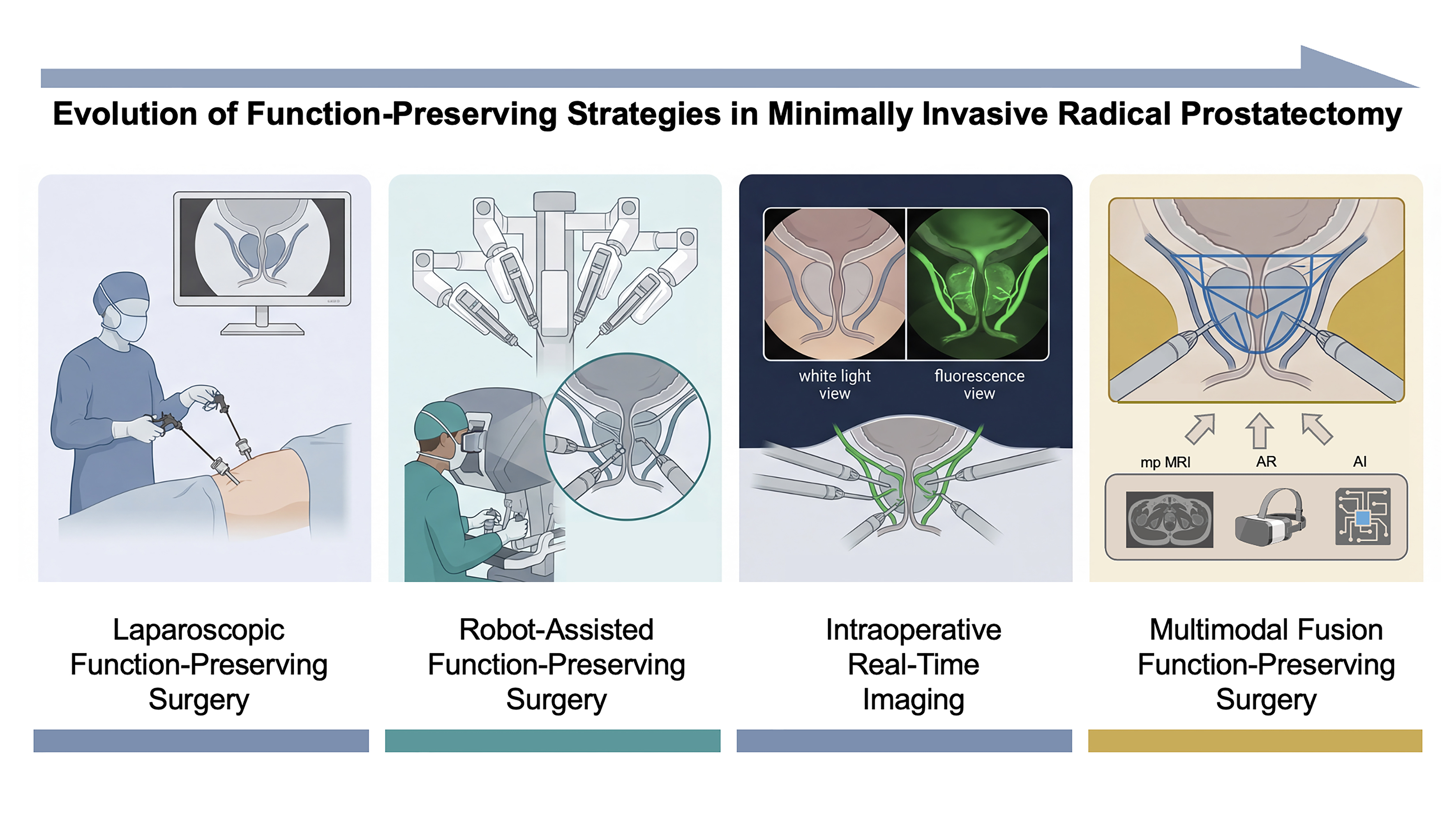
Figure 1. Evolution of function-preserving strategies in minimally invasive radical prostatectomy.
Laparoscopic function-preserving surgery
LRP represents an important stage in the development of minimally invasive surgery for prostate cancer and serves as an important reference for understanding the subsequent advantages of robotic platforms. Its main limitations include a two-dimensional operative view, restricted instrument freedom, and a prolonged learning curve for complex suturing, all of which directly affect the consistency of nerve sparing, urethral reconstruction, and functional recovery.
The broader evidence comparing robotic and open RP also supports a cautious functional advantage for RARP. A 2026 systematic review and meta-analysis of 27 prospective comparative studies involving 38,530 patients associated RARP with less blood loss, fewer transfusions and overall complications, shorter hospitalization, and better early continence and erectile-function recovery than open RP; however, heterogeneity was substantial for several outcomes, and additional randomized trials with longer follow-up remain necessary[8]. For LRP vs. RARP, high-quality direct comparative evidence comes from the LAP-01 randomized, patient-blinded, multicenter trial and its 12-month follow-up. In this trial of 782 patients, RARP was associated with a higher continence recovery rate than LRP at 3 months (54% vs. 46%), whereas the between-group difference attenuated and was no longer statistically significant at 12 months[9,10]. These findings suggest that LRP can achieve function preservation, but its recovery trajectory may depend more heavily on surgeon experience and may be less favorable than RARP with respect to early continence recovery.
A similar pattern is observed for sexual function recovery. A 2025 systematic review and meta-analysis including 13 studies and 6,281 patients showed that RARP was associated with better erectile function recovery than LRP at 3, 6, and 12 months after surgery, with a 12-month risk difference of 0.06 [95% confidence interval (CI), 0.02-0.10][11]. Thus, LRP retains an important place in the evolution of minimally invasive prostate cancer surgery; however, when the primary goals are nerve preservation and accelerated functional recovery, robotic platforms may offer advantages in early recovery.
Robot-assisted function-preserving surgery: multiport and single-port platforms
Multiport robot-assisted radical prostatectomy (MP-RARP) remains the minimally invasive approach supported by the most mature body of evidence and serves as the principal reference platform when discussing nerve-sparing surgery. Its advantages lie not only in 3D high-definition visualization and wristed instruments, but also in the resulting ability to identify anatomical planes more consistently, control traction and thermal injury, and perform more refined urethral reconstruction. Although anterior, lateral, and posterior approaches differ in specific operative steps, they share the same objective: to achieve a better balance between oncological control and function preservation.
On this platform, postoperative functional outcomes appear to depend less on the use of a robot per se than on the specific mode of nerve preservation and tissue handling. Guimarães et al. reported that anterior periprostatic tissue preservation and a clipless technique were associated with shorter mean time to continence recovery (5.8 monthsvs. 6.6 months) and a higher 12-month erectile function recovery rate (75.0% vs. 53.5%)[12]. Görgen et al. further showed that patients with higher NVB preservation scores had a 12-month trifecta rate of 66.6%, compared with 33.3% in the lower-score group[13]. However, nerve sparing is an independent risk factor for ipsilateral positive surgical margins [odds ratio (OR) 1.42][14]. The value of MP-RARP also lies in the development of intraoperative decision-making systems such as the neurovascular structure-adjacent frozen-section examination (NeuroSAFE). The NeuroSAFE PROOF trial demonstrated that NeuroSAFE-guided RARP improved 12-month erectile function [mean International Index of Erectile Function-5 (IIEF-5): 12.7 vs. 9.7] and short-term urinary continence at 3 months[15]. A 2025 systematic review and meta-analysis (4,207 patients) associated NeuroSAFE with higher nerve-sparing rates (OR 5.49), lower positive margins (OR 0.55), lower biochemical recurrence (OR 0.47), and better 12-month functional recovery (ORs 2.01 and 3.50)[16]. Nevertheless, due to the predominance of nonrandomized studies and limited long-term follow-up, these findings remain supportive rather than definitive.
Single-port robot-assisted radical prostatectomy (SP-RARP) can be understood as an optimization of the surgical access route within existing RARP principles rather than a redefinition of nerve preservation. Its main advantages lie in fewer incisions, better postoperative pain control, and shorter hospital stay. A 2025 updated meta-analysis (15 studies, 3,116 patients) found that SP-RARP was associated with lower estimated blood loss, shorter hospital stays, earlier catheter removal, lower pain scores, and less opioid use, whereas continence, potency, complications, positive margins, biochemical recurrence, and operative time did not differ significantly from MP-RARP[17]. A recent propensity-matched comparison [285 single-port (SP) transvesical vs. 285 multiport (MP) transperitoneal] reported higher same-day discharge rates, shorter catheterization duration, less opioid prescribing, and better 3-month continence with the SP approach, whereas 6-month continence, 12-month potency, and oncological outcomes were similar; however, the study was retrospective and single-center[18]. Thus, the most prudent current conclusion is not that SP-RARP has surpassed MP-RARP in long-term outcomes, but rather that SP approaches may improve selected perioperative recovery metrics and possibly early continence in experienced settings without compromising short- to mid-term cancer control. The technical characteristics and reported outcomes of the different approaches are shown in Table 1.
Classification-based comparison of the main RARP access routes and the SP platform for function-preserving radical prostatectomy
| Approach | Initial entry site | Key anatomical plane | Reported clinical contexts | Complications and functional highlights | Oncological expectations |
| Anterior (TP) | Enter the Retzius space first through an anterior peritoneal incision | Early exposure of the endopelvic fascia, puboprostatic/pubovesical ligaments, DVC, bladder neck, and then the posterior plane | Most familiar route for routine localized disease and for surgeons trained in open anatomy; facilitates standard PLND | Provides the largest working space. Hood-type anterior preservation reported continence of 21%, 36%, 83%, 88%, and 91% at 1, 2, 4, 6, and 12 weeks, with a 6% PSM rate in a single-center series[19] | Mature oncological experience; functional recovery depends heavily on how much anterior/periurethral support is preserved and on tumor location[20] |
| Posterior RS (Bocciardi, TP) | Posterior entry via the pouch of Douglas, exposing the vas deferens and seminal vesicles first | Posterior intrafascial dissection between Denonvilliers’ fascia and the rectum; preserves the puboprostatic/pubovesical complex and usually avoids DVC ligation | Reported in settings where early continence recovery is prioritized; requires experienced surgeons and careful anatomical and oncological selection | The main advantage is faster continence recovery. In a randomized study, immediate continence was 51% vs. 21%, with median recovery of 1 dayvs. 21 days; meta-analyses confirm the early continence benefit[21,22] | Short- to mid-term cancer control is generally comparable, but margin caution is warranted in selected anterior or ≥ pT3 disease[21,22] |
| Lateral (TP) | A lateral “buttonhole” is created through the bladder neck-prostate-vascular pedicle/NVB triangle | Posterolateral structures and the NVB are addressed first, while the anterior pubovesical complex is preserved as much as possible | Selected localized cases in which extensive NVB preservation is considered; requires familiarity with posterolateral hemostasis | In a 513-patient series, immediate continence was 85%-86% and 1-year potency 72%-73%[23] | PSM was 32.9%-37.9%, whereas clinically significant margins were 5.9%-7.6%; evidence is mainly retrospective and from high-volume centers[23] |
| VIP (TP) | Anterior transperitoneal access on the robotic platform | Progressive preservation of lateral/posterolateral fascial tissue; veil and super-veil refinements extend anterior fascial and nerve preservation | Used when continence and potency preservation is strongly emphasized in organ-confined disease; requires a highly experienced team | The 2009 VIP technical update formalized superveil nerve sparing and other refinements to improve trifecta-oriented recovery while maintaining the minimally invasive benefits of robotic surgery[24] | Feasible with good cancer control in expert hands, but technical complexity has limited broad dissemination[24] |
| Extraperitoneal | The working space is created outside the peritoneal cavity without entering the abdomen | Standard prostate dissection is completed within the preperitoneal space | Useful when minimizing bowel manipulation is desirable, including in patients with prior abdominal surgery or concern for postoperative ileus | A meta-analysis of 16 studies (3,897 patients) showed faster operative time, shorter stay, and lower postoperative ileus and inguinal hernia rates than transperitoneal RARP[25] | Positive margins and 6-month continence were similar to transperitoneal RARP[25] |
| Transvesical (mainly SP) | Direct entry through the bladder dome, with the prostate approached from within the bladder | Regionalized intravesical dissection limits disturbance of the anterior Retzius structures | Reported as an option for patients with hostile abdomens or prior abdominal surgery, and in settings emphasizing rapid recovery and short catheterization | In a matched comparison, SP transvesical RARP achieved 47% immediate continence and 82.8% continence at 3 months, with shorter catheterization (4 daysvs. 7 days) and markedly less opioid prescribing[18] | Short- to mid-term oncological outcomes were similar to standard MP transperitoneal RARP in experienced centers[18] |
| Perineal route | Perineal access directly beneath the prostate, avoiding the peritoneal cavity | Dissection is performed through the perineal route with limited pelvic working space | May be considered in selected patients with complex abdominal surgical history or frozen pelvis, when abdominal access is problematic | Offers a scar-sparing nonabdominal route, but comparative evidence remains limited and the procedure is technically demanding[26] | In a matched study of SP radical perineal prostatectomy, nonlimited PSMs were higher than with MP transperitoneal RARP, although 1-year biochemical recurrence was similar[26] |
| SP platform | Single skin incision using the purpose-built SP robotic platform; can be deployed transperitoneally, extraperitoneally, transvesically, or transperineally | “Regionalized” access with multi-jointed instruments and fewer abdominal wall punctures | Use has mainly been reported in centers with platform-specific experience when smaller access trauma and accelerated discharge are priorities | An updated meta-analysis showed less blood loss, shorter hospital stay, earlier catheter removal, and lower pain/opioid use than MP-RARP, while continence and potency were broadly similar overall[17] | Short- to mid-term oncological outcomes appear comparable to MP-RARP, but long-term and approach-specific evidence remains less mature[17,18] |
Intraoperative fluorescence imaging-assisted function-preserving surgery
If refinements of robotic dissection focus on how tissue is handled, intraoperative fluorescence imaging addresses how key structures can be visualized in real time. The most widely studied strategy in RP uses intravenous indocyanine green (ICG) with near-infrared fluorescence (NIRF) integrated into the robotic platform to enhance visualization of periprostatic vessels, the landmark artery within the NVB, and local perfusion. In principle, this may help the surgeon identify safer dissection planes, limit unnecessary traction or cautery near the NVB, and improve hemostasis, thereby supporting postoperative functional recovery. At present, however, the evidence base remains limited and is derived mainly from feasibility and early clinical studies rather than randomized comparative trials.
In the initial clinical experience reported by Mangano et al., indocyanine green-near-infrared fluorescence (ICG-NIRF) was used during nerve-sparing RARP to identify the landmark artery and facilitate preservation of the NVB, demonstrating technical feasibility and satisfactory intraoperative visualization[27]. More recently, Amara et al. analyzed 91 consecutive patients with localized prostate cancer who underwent ICG-NIRF-guided RARP and reported successful NVB identification in all cases, no ICG-related complications, and no significant difference between baseline and 9-month Sexual Health Inventory for Men (SHIM) scores (P = 0.331), with erectile function returning to baseline in most patients[28]. These findings suggest that fluorescence guidance may be a useful adjunct for real-time microanatomical dissection. Nevertheless, because current studies are mostly retrospective, non-comparative, and focused on short-term feasibility, intraoperative fluorescence imaging should presently be regarded as a promising supportive tool rather than an established standard for function preservation.
Multimodal fusion function-preserving surgery
While fluorescence imaging improves what can be seen intraoperatively, multimodal data fusion and 3D reconstruction seek to improve what can be understood before and during surgery. In current practice, preoperative multiparametric magnetic resonance imaging (mpMRI) remains the core imaging substrate, but image-derived information can also be integrated with biopsy, clinical, and perioperative variables to support both anatomical navigation and side-specific risk assessment. By segmenting the prostate, index lesion, capsule, urethra, and adjacent NVBs from preoperative imaging into patient-specific 3D models, surgeons can translate tumor location and extracapsular extension risk into a more intuitive operative map. When these models are used for cognitive review or overlaid onto the robotic console as AR, the goal is not to replace intraoperative judgment, but to make nerve-sparing decisions more individualized and anatomically explicit.
Schiavina et al. overlaid preoperative augmented-reality 3D models onto the intraoperative field in 26 RARP cases and found that the nerve-sparing plan changed in 38.5% of patients and 34.6% of sides, with overall system applicability of 94.4%; on a 32-region map, lesion-localization sensitivity, specificity, and accuracy were 70%, 100%, and 92%, respectively[29]. In a later randomized clinical trial secondary analysis including 92 patients, Shirk et al. reported that use of 3D digital models for planning robotic prostatectomy was associated with lower 18-month biochemical recurrence (0% vs. 17.9%), less adjuvant or salvage radiotherapy (3.1% vs. 31.6%), better sexual function (mean SHIM score 16.8 vs. 9.8), and a higher trifecta rate (48.0% vs. 10.0%), while continence was similar between groups[30].
The prospective multicenter RIDERS randomized trial provides more direct evidence. Among 133 patients with extracapsular extension or bulging on preoperative magnetic resonance imaging (MRI) who underwent nerve-sparing RARP, 3D-AI-AR guidance was associated with a lower overall positive surgical margin (PSM) rate (22% vs. 39%), less postoperative radiotherapy (18% vs. 35%), and higher 12-month zero-pad continence (91% vs. 71%) than cognitive MRI guidance, while potency and short-term biochemical recurrence were similar[31]. These results are encouraging, but they arise from a selected population and 12 months of follow-up; longer-term oncological assessment and external replication are required before routine implementation.
FUNCTION-PRESERVING FOCAL ABLATION FOR PROSTATE CANCER
Current evidence suggests that focal ablation may be most appropriate for carefully selected patients with low- to intermediate-risk disease in whom lesions can be adequately delineated by imaging and targeted biopsy. Focal therapy is not yet a standard option, but it may be discussed during shared decision-making with carefully selected, fully informed patients[32]. EAU guidance places particular emphasis on prospective evidence generation, with registry-based recommendations for high-intensity focused ultrasound (HIFU) or cryotherapy and trial-based recommendations for other ablative modalities[2]. These recommendations coexist with current clinical practice: some focal modalities are already used in selected clinical settings, including outside formal trials where locally permitted, although their availability and pathways for implementation vary by jurisdiction. Accordingly, the evaluation of focal therapy in this review is framed around functional benefit, patient selection, surveillance, and the maturity of oncological evidence, rather than as evidence of equivalence to established curative standards. Figure 2 illustrates currently common focal ablation techniques and their underlying principles.
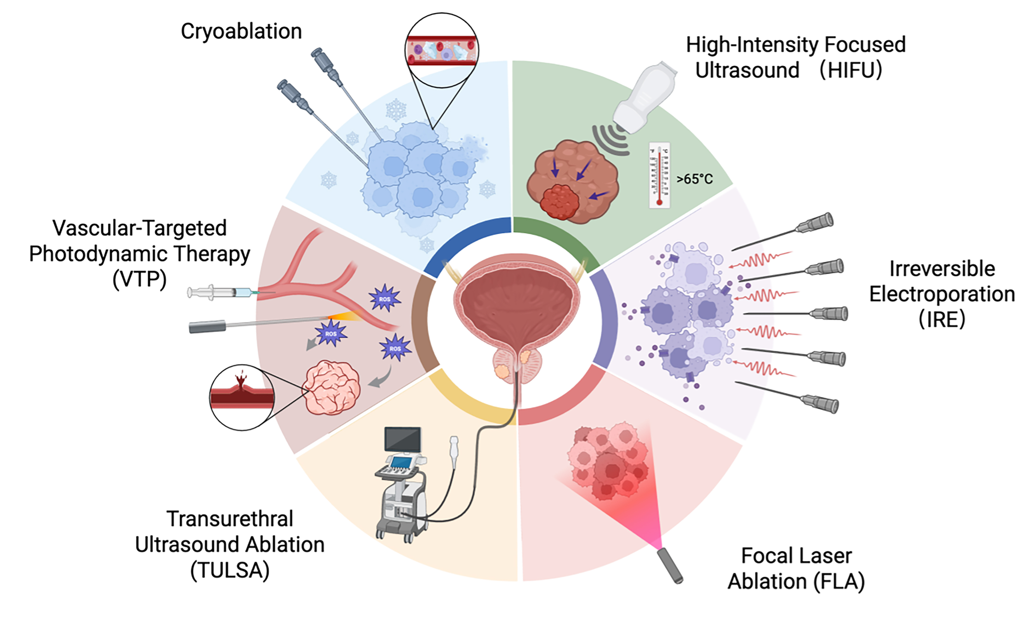
Figure 2. Schematic illustration of the major focal ablation techniques for prostate cancer and their mechanisms of action. [Created in BioRender. Zhang, C. H. (2026) https://BioRender.com/hda0tdq]. VTP: Vascular-targeted photodynamic therapy; TULSA: transurethral ultrasound ablation; FLA: focal laser ablation; IRE: irreversible electroporation; HIFU: high-intensity focused ultrasound.
Cryotherapy
Cryotherapy induces tissue necrosis through intracellular ice crystal formation, membrane disruption, and microvascular thrombosis caused by low temperature. Its major advantage is that the ablation boundary can be monitored under ultrasound guidance, while temperature probes can help protect the urethra and rectum. Its principal appeal in focal therapy therefore lies in perioperative safety and the potential for function preservation; long-term oncological durability has not been conclusively established. An early systematic review reported post-focal cryotherapy continence rates of 96%-100%, urinary retention rates of 0%-15%, and a very low rate of rectourethral fistula, while erectile dysfunction ranged widely from 0% to 42%[33].
Because of its relatively low perioperative morbidity, cryotherapy has long been considered a tissue-preserving option for carefully selected patients. However, its key limitation lies not in short-term safety but in the continued insufficiency of long-term oncological evidence. In the studies included in the cited systematic review, the positive follow-up biopsy rate was 21.8%[33]. Cryotherapy may therefore be considered a function-preserving option in selected patients, but current evidence does not establish equivalence to RP or radiotherapy.
HIFU
HIFU uses focused ultrasound to generate high temperature within the target zone, leading to protein coagulation and cell death, and is one of the most widely applied focal therapies with a relatively substantial evidence base. Thus, the assessment of HIFU should not center solely on theoretical feasibility, but on whether it appears to have developed relatively stable mid-term oncological control and functional outcomes. In a multicenter prospective cohort reported by Guillaumier et al., 625 patients with clinically significant, nonmetastatic prostate cancer were included; the 5-year failure-free survival (FFS) was 88%, metastasis-free survival (MFS) 98%, cancer-specific survival (CSS) 100%, and OS 99%. Among patients completing questionnaires, 241/247 (98%) maintained complete pad-free continence, and only 2% used any pads[34].
However, the outcomes of HIFU are still clearly influenced by case selection and treatment extent. A 2024 systematic review of whole-gland HIFU that included 6,618 patients reported biochemical disease-free survival rates ranging from 21.7% to 89.2% and negative biopsy rates ranging from 20% to 92.7%, indicating substantial inter-center variability[35]. These whole-gland results cannot be directly extrapolated to focal HIFU. Overall, HIFU may currently be regarded as one of the focal ablative modalities supported by a relatively more substantial evidence base, but its clinical use still depends on strict patient selection and standardized follow-up.
Irreversible electroporation
Irreversible electroporation (IRE) is a nonthermal ablation technique that induces apoptosis by creating irreversible nanopores in the cell membrane through high-voltage pulses. Because it causes relatively limited damage to collagen scaffolds and surrounding heat-sensitive structures, IRE is theoretically well suited to preserving the urethra, sphincter, and NVBs; accordingly, its most closely watched advantage relates to functional preservation.
Available clinical data largely revolve around this point. The prospective Targeted Ablation Using Ultrasound-Guided Irreversible Electroporation of Index Tumors (TARGET) study enrolled 20 intermediate-risk patients, 19 of whom completed treatment. At 6 months, urinary and sexual function scores showed no clear decline; at 12 months, 14/19 (74%) patients had no clinically significant cancer detected throughout the whole gland; and the 2- and 4-year rates of freedom from radical treatment were 79% and 73%, respectively[36]. In addition, in a long-term cohort of 229 patients reported by Scheltema et al., the median follow-up was 60 months, the 5-year FFS was 84%, MFS was 99.6%, and both CSS and OS were 100%; continence at 12 months was maintained from 98% at baseline to 99%, but erections sufficient for intercourse declined from 71% to 58%[37].
More recently, the prospective, nonrandomized, multicenter PRESERVE trial treated 121 patients with organ-confined Grade Group 2-3 intermediate-risk disease. At 12 months, the primary intention-to-treat negative in-field biopsy rate for any Gleason-gradable cancer was 71% (86/121; 95%CI: 62%-79%); using the Delphi criterion for absence of clinically significant cancer, the negative in-field rate was 84% (95%CI: 76%-90%). Urinary scores changed little on average, and 84% of patients with good baseline sexual function maintained erections sufficient for penetration. Fourteen patients (12%) experienced grade ≥ 3 adverse events, including three procedure-related grade 3 events[38]. Its single-arm design and 12-month follow-up limit direct comparison with longer-term cohort estimates of FFS or residual disease.
Therefore, data on continence preservation with IRE are relatively stable, but sexual function is not completely unaffected, and residual local disease still exists. At present, the most appropriate interpretation is that IRE has demonstrated tissue-preserving potential in selected cohorts, but high-quality comparative trials are still lacking to show that it is superior to other focal modalities.
Transurethral ultrasound ablation
Transurethral ultrasound ablation (TULSA) delivers outward circumferential ultrasound from a transurethral applicator and, in combination with real-time MRI thermometry, enables whole-gland or partial-gland ablation. Unlike the transperineal needle-based approach, its technical characteristic is a more controllable treatment path, with dynamic adjustment of energy deposition according to the real-time thermal map; thus, in theory, it may offer both precision and safety.
At present, this advantage is reflected mainly in early functional outcomes. A systematic review including 10 studies and 224 patients showed retreatment rates of 7%-17% after a single TULSA session, continence preservation rates of 92%-100%, erectile function preservation rates of 75%-98%, and a 6% rate of grade III adverse events, with no rectal injury or grade IV complications reported[39]. These findings suggest that TULSA has substantial promise from a functional perspective, although current evidence still comes mainly from early single-arm studies and its long-term oncological role requires cautious interpretation.
Focal laser ablation
Focal laser ablation (FLA) delivers thermal energy precisely to the target lesion through optical fibers and has mainly been evaluated in smaller, well-demarcated lesions visible on MRI. Because the treatment field is limited and the access route is relatively precise, FLA is often regarded as a minimally invasive focal technique. Published FLA cohorts have included heterogeneous populations spanning low- to intermediate-risk disease, precluding definition of a single universal indication.
However, current evidence suggests that the main issue with FLA is not short-term feasibility, but rather fragmented research and nonuniform outcome assessment. A 2026 systematic review and meta-analysis including 12 studies and 421 patients reported a 15% recurrence rate of clinically significant cancer and a mean prostate-specific antigen (PSA) decline of 2.3 ng/mL at 12 months; although sexual function declined statistically, the effect size was small, and urinary function showed no significant change[40]. Another 2024 systematic review and network meta-analysis of MRI-guided FLA included 9 trials and 296 patients, and reported a residual lesion rate of 20.37%, a cancer-free survival rate of 75.62%, and a combined rate of major and minor complications of 14.26%[41]. Thus, based on current data, the advantages of FLA appear to lie in low invasiveness and limited functional impairment, but the absence of a standardized reassessment pathway and of high-quality comparative studies remains a key barrier to its broader adoption.
Vascular-targeted photodynamic therapy
Vascular-targeted photodynamic therapy (VTP) involves intravenous administration of a photosensitizer followed by activation with laser light of a specific wavelength, thereby inducing destruction of the tumor-feeding vasculature. Unlike thermal ablation, its potential advantage lies in the possibility of relatively selective treatment of the targeted region, with comparatively limited direct injury to the urethral sphincter and NVBs; it is therefore particularly relevant when asking whether control and function preservation can be balanced within a narrow set of indications.
The most persuasive data for VTP currently come from a phase III randomized controlled trial. Azzouzi et al. randomized 413 men with low-risk localized prostate cancer to VTP or active surveillance. At 24 months, disease progression rates were 28% and 58%, respectively, while negative biopsy rates were 49% and 14%; grade 3-4 adverse events were infrequent, and although urinary retention was relatively common, it usually resolved within 2 months[42]. Thus, randomized evidence for VTP in low-risk localized disease is relatively clear, but its conclusions apply mainly to a narrowly defined indication and cannot be generalized to a broader population with localized prostate cancer. Table 2 provides a descriptive summary of representative studies stratified by ablative modality.
Descriptive summary of representative clinical studies of major focal ablation techniques
| Modality | Study | Number of patients | Patient risk group | Efficacy outcomes | Complications/Functional outcomes |
| Cryotherapy | Shah 2019 Prospective multicenter registry[43] | 122; median follow-up 27.8 months | Nonmetastatic clinically significant PCa; NCCN intermediate risk 71.3%, high risk 28.7% | 3-year FFS 90.5%; 93.3% in intermediate-risk and 84.7% in high-risk disease | 0% required pads; ED 16.1% (5/31) |
| Shah 2021 Functional recovery analysis[44] | 58 (PROM subgroup of the 122-patient cohort) | Predominantly Gleason 3+4/4+3 and cT2 disease | Return to baseline IPSS: 78% at 12 months, 87% at 18-24 months; return to baseline IIEF-EF: 85% at 12 months, 89% at 18-24 months | Urinary and sexual function recovery were the primary endpoints; high-grade complications were not reported separately | |
| Tay 2016 Systematic review[33] | 272 mandatory re-biopsies; functional data from 1,582 patients | Included studies mainly involved localized PCa with heterogeneous risk stratification | Positive biopsy rate on follow-up 21.8% | Continence 96%-100%; ED 0%-42%; urinary retention 0%-15%; rectourethral fistula 0%-0.1% | |
| HIFU | Rischmann 2017 Prospective multicenter hemiablation study[45] | 111 | Unilateral, localized disease, predominantly favorable-risk | 1-year freedom from clinically significant cancer 95%; 2-year freedom from radical treatment 89% | 12-month continence 97%; erectile function preservation 78%; grade 3 adverse events 13% |
| Guillaumier 2018 Multicenter 5-year cohort[34] | 625; median follow-up 56 months | Clinically significant, nonmetastatic PCa; 84% intermediate/high risk | 5-year FFS 88%; MFS 98%; CSS 100%; OS 99% | 241/247 (98%) completely pad-free; any pad use 2% | |
| Shoji 2025 Multicenter prospective study[46] | 240; median follow-up 48 months | D’Amico low risk 51, intermediate risk 107, high risk 82 | Biochemical DFS after single treatment: 93.7%/88.5%/84.8% (low/intermediate/high risk); pathological DFS: 92.2%/91.6%/86.6% | Urinary and sexual function declined transiently at 1 month and returned to baseline by 3-6 months | |
| IRE | Blazevski 2020 Prospective biopsy-monitored cohort[47] | 123 | Localized clinically significant PCa | No residual lesion on follow-up biopsy in 90.2%-97.3%; 3-year avoidance of whole-gland treatment 96.75% | Minimal overall impact on quality of life |
| TARGET study (Fainberg 2024)[36] | 20 enrolled, 19 treated | Intermediate-risk PCa | No clinically significant cancer in the whole gland at 12 months in 74%; 2- and 4-year freedom from radical treatment 79% and 73% | No clear decline in urinary or sexual function scores at 6 months | |
| Scheltema 2023 5-year cohort[37] | 229; median follow-up 60 months | 86% intermediate risk, 7% high risk | FFS: 91% at 3 years, 84% at 5 years, and 69% at 8 years; MFS 99.6%; CSS/OS 100% | Continence 98% → 99%; erections sufficient for intercourse 71% → 58%; residual csPCa on follow-up biopsy 24% | |
| IRE | George 2026 PRESERVE prospective multicenter single-arm trial[38] | 121; 12 months | Organ-confined GG2-3 intermediate-risk disease; cT1-T2c | 12-month negative in-field biopsy: any Gleason-gradable cancer 71% (86/121); no csPCa by Delphi criterion 84% | Small mean urinary-score changes; 84% with good baseline sexual functionmaintained penetration-sufficient erections; grade ≥ 3 adverse events 12% (14/121), including 3 procedure-related events |
| TULSA | Dora 2022 Systematic review[39] | 10 studies, 224 patients | Localized PCa; heterogeneous inclusion criteria | Retreatment rate 7%-17% | Continence 92%-100%; erectile function preservation 75%-98%; grade III adverse events 6%; no rectal fistula or grade IV complications |
| Nair 2021 Prospective phase I study, 3-year outcomes[48] | 30 | Localized PCa; predominantly low risk | csPCa in 10/29 (34%) at 3 years; PSA nadir 0.33 ng/mL, approximately 0.8 ng/mL at 3 years | No new severe complications between years 1 and 3; urinary and bowel function stable; erectile function recovered by 1 year and was maintained | |
| Klotz 2021 Prospective TACT study[49] | 115 | Low- to intermediate-risk; GG2 63%, NCCN intermediate risk 67% | PSA reduction ≥ 75% in 96% (110/115); no cancer on 12-month biopsy in 65% (72/111) | Grade 3 adverse events 8%; 96% returned to baseline urinary continence; 75% of potent men maintained or regained penetration-sufficient erections | |
| FLA | Bushati 2026 Systematic review/meta-analysis[40] | 12 studies, 421 patients | Localized PCa with heterogeneous risk stratification | Clinically significant cancer recurrence 15%; mean PSA decline 2.3 ng/mL at 12 months | Mild decline in sexual function; no significant change in urinary function |
| Marcelin 2024 Systematic review/network meta-analysis[41] | 9 trials, 296 patients | Mainly MRI-visible lesions | Residual lesion rate 20.37%; cancer-free survival 75.62% | Combined rate of major and minor complications 14.26% | |
| Eggener 2016 Phase II study[50] | 27 | Highly selected localized PCa | Negative biopsy in the ablation zone in 26/27 (96%) at 3 months; favorable 1-year oncological outcomes | Acceptable urinary, erectile, and bowel toxicity | |
| Walser 2019 Cohort of 120 patients[51] | 120 | Low- to intermediate-risk disease | Freedom from retreatment at 1 year 83%; median PSA reduced to 3.25 ng/mL at 12 months | No significant decline in urinary or sexual function; mild hematuria common; 2 early rectourethral fistulas | |
| Chao 2021 5-year follow-up[52] | 36 enrolled, 30 evaluable; median follow-up 71 months | cT1c-T2a, PSA < 10, GG < 4 | 5-year FFS 83%; local recurrence 40%; 2 metastases; no PCa deaths | Repeat focal or whole-gland salvage treatment was relatively common | |
| VTP | Taneja 2016 Phase I/II multicenter study[53] | 30 | Unilateral, low-volume, Gleason 3+3 low-risk disease | When optimized dose plus LDI ≥ 1 was achieved, the negative biopsy rate in the treated lobe was 73.3% | Minimal impact on urinary function, sexual function, and overall quality of life |
| Azzouzi 2017 Phase III randomized controlled trial[42] | 413; 24-month follow-up | Low-risk localized PCa | Disease progression at 24 months: 28% vs. 58%; negative biopsy 49% vs. 14% (control: active surveillance) | Few grade 3-4 adverse events; urinary retention was more common but usually resolved within 2 months |
Available cross-study data support descriptive synthesis but not reliable comparative ranking. A 2025 systematic review and meta-analysis of 49 cohorts found no significant between-modality differences in pooled outcomes for focal cryotherapy, HIFU, and IRE; however, these were indirect cross-study comparisons limited by substantial heterogeneity and did not constitute head-to-head evidence[54]. Cryotherapy and HIFU have broader published experience and often report favorable continence outcomes. IRE and TULSA have reported relatively favorable urinary and sexual functional outcomes in selected cohorts, but current evidence does not establish that they provide superior protection of sexual function. FLA studies include heterogeneous risk groups, including low- to intermediate-risk disease, whereas the pivotal randomized VTP evidence was restricted to a low-risk population. FLA and VTP have distinct evidence bases, and a single pooled 5-year residual or recurrence estimate cannot be derived from studies using incompatible endpoints and follow-up durations.
Treatment selection should be individualized through multidisciplinary and shared decision-making that considers tumor risk, lesion distribution, life expectancy, baseline function, patient preferences, and willingness to undergo repeat biopsy, retreatment, or salvage therapy. For higher-risk or multifocal localized disease, guideline-concordant management may include RP as part of a multimodal strategy or radiotherapy with systemic therapy when indicated. Within this shared decision-making process, focal therapy may be discussed with carefully selected, fully informed patients, although it is not yet a standard option[32]; some focal modalities are already used in selected clinical settings, including outside formal trials where locally permitted. Counseling should address limitations in comparative and long-term evidence, structured surveillance, and possible repeat or salvage treatment; availability, regulation, reimbursement, and implementation remain jurisdiction-specific.
AI-ASSISTED SURGERY
With the continued refinement of minimally invasive RP and focal ablative techniques, a central challenge in function-preserving treatment is not simply whether a procedure can be performed, but how accurately surgeons can select candidates, individualize nerve-sparing or ablative strategies, and anticipate both oncological and functional outcomes. AI is increasingly being explored as an adjunct across the surgical workflow, including preoperative risk stratification, MRI-based segmentation and 3D planning, intraoperative navigation, outcome prediction, and workflow optimization. Most evidence remains concentrated in diagnostic imaging, digital pathology, and retrospective prognostic modeling, although the RIDERS trial provides early randomized evidence for 3D-AI-AR guidance during selected nerve-sparing RARP[31,55]. AI should therefore be regarded as an emerging decision-support and guidance technology rather than an established intraoperative standard for function preservation.
Preoperative risk stratification and surgical decision support
In function-preserving surgery, decisions regarding whether and to what extent the NVB should be preserved, which surgical access route should be selected, and whether focal ablation is appropriate depend on accurate assessment of tumor aggressiveness, extracapsular extension, lymph node involvement, and patient-specific competing risks. AI models may contribute by integrating clinical, imaging, biopsy, and pathological variables into individualized risk estimates. A systematic review comparing MRI-based AI methods with conventional clinical assessment showed higher areas under the summary receiver operating characteristic curves (SROC-AUCs) for AI in clinically significant prostate cancer detection (0.87 vs. 0.82) and adverse pathology prediction (0.86 vs. 0.75), although the authors emphasized the need for prospective validation and open testing datasets[56].
Several clinical decision-support models further illustrate this potential, but their interpretation should remain cautious. A study based on data from 118,236 patients with localized prostate cancer in the Surveillance, Epidemiology, and End Results (SEER) Program developed a final treatment recommendation model to estimate survival probabilities after surgery or radiotherapy; the radiotherapy and surgery models achieved C-index ranges of 0.735-0.787 and 0.769-0.797, respectively, and patients whose actual treatment was consistent with the model recommendation had higher survival rates[57]. Another SEER-based model using the CatBoost gradient-boosting algorithm and SHapley Additive exPlanations (SHAP) for model interpretation identified T stage, disease stage, age, positive core rate, PSA, and Gleason score as key decision variables, with the 10-year survival rate in patients undergoing RP reported as 89.2%[58]. In a more surgery-specific setting, the Dr. Answer AI clinical decision-support system was developed from 7,128 patients treated with RP and predicted tumor-node-metastasis (TNM) stage, extracapsular extension, seminal vesicle invasion, and lymph node metastasis; the random forest model achieved a 76.98% recall for TNM staging, whereas the k-nearest neighbor model achieved recalls of 80.24%, 98.67%, and 95.45% for extracapsular extension, seminal vesicle invasion, and lymph node metastasis, respectively[59]. These models may support shared decision-making, but they should not be interpreted as replacing guideline-based multidisciplinary assessment.
Image segmentation and 3D surgical planning
Accurate preoperative planning is a prerequisite for individualized nerve-sparing and focal therapy. Deep learning-based prostate MRI analysis has been applied to prostate gland segmentation, lesion detection, volume estimation, and characterization of tumor aggressiveness, thereby providing structured inputs for patient-specific 3D models[60]. In principle, these tools can help delineate the prostate, index lesion, capsule, urethra, and adjacent structures, and can facilitate MRI-ultrasound or MRI-histopathology registration for treatment planning[61]. For function-preserving surgery, the most clinically relevant application is to convert tumor location and side-specific extracapsular extension risk into a clearer surgical map. Nevertheless, automatic identification of fine structures such as the NVB remains technically challenging, and AI-derived 3D planning should be considered complementary to surgeon interpretation, high-quality mpMRI review, and intraoperative judgment.
Intraoperative navigation and real-time assistance
The role of AI in true intraoperative guidance for prostate cancer surgery remains developmental. The RIDERS randomized trial reported lower overall PSMs, less postoperative radiotherapy, and higher 12-month zero-pad continence with 3D-AI-AR guidance than with cognitive MRI guidance, while potency and short-term biochemical recurrence were similar[31]. These findings constitute direct clinical evidence relevant to margin control and continence, but the trial enrolled 133 selected patients with extracapsular extension or bulging on MRI, follow-up was limited to 12 months, and longer-term replication is needed. Beyond this trial, evidence for surgical step recognition, anatomical landmark detection, instrument tracking, bleeding detection, and automated image registration is derived predominantly from retrospective, feasibility, or technical studies and reviews[62]. Evidence that these applications improve nerve preservation or functional outcomes in routine RARP remains insufficient. AI-based prostate-specific membrane antigen positron emission tomography/computed tomography (PSMA PET/CT) segmentation tools may standardize lesion identification and reporting; for example, the aPROMISE platform was evaluated against a standard image viewer and histopathology in patients undergoing PSMA-radioguided surgery[63]. This evidence supports structured preoperative or perioperative imaging interpretation rather than direct robotic-console navigation.
The integration of fluorescence imaging with AI should also be regarded as a future direction rather than an established clinical standard. ICG-NIRF can visualize periprostatic vascular structures and the landmark artery within the NVB, as discussed above, while AI-based image analysis could theoretically enhance vessel recognition and decision support. However, prospective studies are still needed to determine whether AI-enhanced fluorescence guidance improves functional outcomes beyond expert visual interpretation.
Prediction of functional outcomes and prognostic assessment
Direct prediction of postoperative functional recovery has been investigated, but the evidence remains exploratory. In 100 RARP cases, a deep-learning survival model combining automated surgical performance metrics with clinical features predicted continence recovery with a C-index of 0.60[64]. A single-center study of 101 patients combined intraoperative video snapshots with ensemble machine learning and reported an internally validated area under the curve (AUC) of 0.882 for early continence recovery[65]. A larger referral-center cohort of 8,524 patients developed artificial neural-network models with AUCs of 0.68 for 12-month continence and 0.74 for potency[66]. These studies demonstrate feasibility but differ in endpoints and inputs, and prospective multicenter external validation and evidence of clinical benefit are still limited.
AI has shown increasing value in oncological prognostication, particularly when digital pathology is combined with clinical variables. A digital pathology-based multimodal AI model externally validated using four phase III trial datasets from the Systemic Therapy in Advancing or Metastatic Prostate Cancer: Evaluation of Drug Efficacy (STAMPEDE) platform was prognostic for prostate cancer-specific mortality in men with advanced or high-risk nonmetastatic prostate cancer starting long-term androgen-deprivation therapy[67]. In metastatic hormone-sensitive prostate cancer, a related digital pathology-based AI biomarker was associated with OS, clinical progression, and progression to castration-resistant disease[68]. These studies support AI-based prognostication in their source populations, but their relevance to localized nerve-sparing RARP and postoperative functional or margin outcomes remains unvalidated.
For postirradiation recurrence assessment, Marinkovic et al. evaluated machine-learning classifiers in 109 patients treated with external-beam radiotherapy, with or without androgen-deprivation therapy, who were assessed after PSA elevation. The Naive Bayes model achieved 100.0% sensitivity, 92.3% specificity, 94.4% positive predictive value, 100% negative predictive value, and 96.7% predictive accuracy for disease progression[69]. These findings concern postirradiation recurrence and do not establish postoperative predictive performance after RP. In pathology, a systematic review of 80 studies found that most AI algorithms addressed biopsy specimens and achieved good to excellent performance for cancer detection and grading; some studies also linked AI-derived histological features with biochemical recurrence, extraprostatic extension, perineural invasion, and disease-free survival[70]. A separate systematic review of AI for early prostate cancer detection reported potential improvements in diagnostic performance and reporting time[71]. Post-RARP functional prediction has instead been investigated in the dedicated studies summarized above[64-66], although prospective multicenter external validation remains insufficient.
Data standardization, validation, regulation, and clinical implementation
Several barriers limit the immediate translation of AI into routine surgical decision-making. First, many studies are retrospective and single-center, with heterogeneous imaging protocols, scanner platforms, histological staining procedures, and annotation standards, all of which can reduce generalizability[72]. Second, robust external validation remains essential. The Prostate Imaging: Cancer AI (PI-CAI) international paired confirmatory study showed that an AI system achieved a higher area under the receiver operating characteristic curve (AUROC) than the pooled performance of 62 radiologists using Prostate Imaging Reporting and Data System version 2.1 (PI-RADS v2.1) for clinically significant cancer detection (0.91 vs. 0.86), but non-inferiority to multidisciplinary routine standard-of-care readings was not confirmed because specificity was slightly lower; the authors therefore emphasized prospective validation before clinical implementation[73].
Regulatory and workflow issues also require caution. Although AI-enabled prostate pathology tools have entered clinical use, regulatory authorization generally applies to adjunctive or secondary review rather than autonomous diagnosis[74]. For example, the U.S. Food and Drug Administration (FDA) De Novo summary for Paige Prostate describes an adjunctive software device intended to assist pathologists in detecting areas suspicious for cancer in prostate biopsy whole-slide images after initial review[75]. Broader implementation will require transparent reporting, continuous performance monitoring, interoperability with picture archiving, pathology, and electronic health record systems, and clear allocation of legal responsibility[76,77]. Explainable AI may partly address the black-box problem: in PI-RADS 3 lesions, an explainable deep learning model improved nonexpert reader confidence and reduced reading time by 58 seconds[78]. Overall, the most appropriate current model is clinician-led human-AI collaboration, in which AI assists risk estimation, visualization, and workflow efficiency while the final surgical and diagnostic decisions remain with the treating team.
CONCLUSION
In summary, current evidence supports a consistent direction for function-preserving treatment of localized prostate cancer: reducing urinary and sexual morbidity should be pursued alongside oncological safety rather than as an independent endpoint. Among radical strategies, minimally invasive RP remains an established curative framework, and MP-RARP currently has the most mature evidence base for balancing cancer control and functional preservation. However, the apparent advantages of specific platforms, dissection routes, or adjunctive technologies must be interpreted in light of patient selection, surgeon experience, endpoint definitions, and follow-up duration. Early randomized evidence for 3D-AI-AR guidance is encouraging, but other AI applications remain predominantly exploratory and require prospective external validation.
For carefully selected patients with localized disease, focal ablative modalities provide additional function-preserving options. Reported benefits include lower treatment burden and favorable continence outcomes in selected cohorts, whereas the degree and durability of sexual function preservation and long-term oncological control remain variable. The available heterogeneous evidence neither establishes superiority among focal modalities nor supports routine substitution of focal therapy for guideline-concordant RP, radiotherapy, or active surveillance. Selection should be individualized through multidisciplinary shared decision-making with carefully selected, fully informed patients. Focal therapy is not yet a standard option, but some focal modalities are already used in selected clinical settings, including outside formal trials where locally permitted. Counseling should address limited long-term evidence, structured surveillance, and possible repeat or salvage treatment. EAU guidance continues to emphasize prospective evidence generation through modality-specific registry and trial recommendations, while implementation remains jurisdiction-specific. Future research should refine nerve-sparing, image-guided navigation, AI-assisted decision support, and functional reconstruction while standardizing endpoints and follow-up.
DECLARATIONS
Acknowledgments
The Graphical Abstract was created in BioRender. Zhang, C. H. (2026) https://BioRender.com/somlfea.
Authors’ contributions
Conception and design: Tang SR, Zhang CH, Zheng ZC
Overall framework and conceptual direction of the review: Pan JH
Manuscript writing: Tang SR
Development and organization of the clinical content and critical revision of the manuscript: Tricard T, Chen MY
Supervision: Zhang CH, Zheng ZC, Tricard T, Chen MY, Pan JH
All authors have read and approved the final version of the manuscript and agree to be accountable for all aspects of the work.
Availability of data and materials
Not applicable.
AI and AI-assisted tools statement
Not applicable.
Financial support and sponsorship
This research was funded by the National Natural Science Foundation of China (Nos. 82373358 and 82573527) and the National Key R&D Program “Intergovernmental International Science and Technology Innovation Cooperation” Key Specialized Program: Researchers Exchange Project (No. 2026YFE0158400).
Conflicts of interest
All authors declared that there are no conflicts of interest.
Ethical approval and consent to participate
Not applicable.
Consent for publication
Not applicable.
Copyright
© The Author(s) 2026.
REFERENCES
1. Bray F, Laversanne M, Sung H, et al. Global cancer statistics 2022: GLOBOCAN estimates of incidence and mortality worldwide for 36 cancers in 185 countries. CA Cancer J Clin. 2024;74:229-63.
2. Cornford P, Tilki D, van den Bergh RCN, et al. Prostate cancer. EAU-EANM-ESTRO-ESUR-ISUP-SIOG guidelines. Arnhem, The Netherlands: EAU Guidelines Office; 2026. ISBN: 978-94-92671-32-5. Available from: https://uroweb.org/guidelines/prostate-cancer [Last accessed on 21 Sep 2026].
3. Bill-Axelson A, Holmberg L, Garmo H, et al. Radical prostatectomy or watchful waiting in prostate cancer - 29-year follow-up. N Engl J Med. 2018;379:2319-29.
4. Czarnogórski MC, Settaf-Cherif L, Koper K, et al. Nerve-sparing techniques in robot-assisted radical prostatectomy - anatomical approach. Expert Rev Med Devices. 2024;21:1101-10.
5. Carbin DD, Shah A, Kusuma VRM. Artificial intelligence in robot-assisted radical prostatectomy: where do we stand today? J Robot Surg. 2024;18:404.
6. Della Corte M, Quarà A, De Cillis S, et al. 3D virtual models and augmented reality for radical prostatectomy: a narrative review. Chin Clin Oncol. 2024;13:56.
7. Sarhan K, Khan N, Prezzi D, et al. Reduction of surgical complications via 3D models during robotic assisted radical prostatectomy: review of current evidence and meta-analysis. J Robot Surg. 2024;18:304.
8. Suartz CV, Costa MSS, Matalani CFA, et al. Comparative functional, perioperative and oncological outcomes of robot-assisted and open radical prostatectomy: a systematic review and meta-analysis. Int Urol Nephrol. 2026; Epub ahead of print.
9. Stolzenburg JU, Holze S, Neuhaus P, et al. Robotic-assisted versus laparoscopic surgery: outcomes from the first multicentre, randomised, patient-blinded controlled trial in radical prostatectomy (LAP-01). Eur Urol. 2021;79:750-9.
10. Stolzenburg J, Holze S, Arthanareeswaran V, et al. Robotic-assisted versus laparoscopic radical prostatectomy: 12-month outcomes of the multicentre randomised controlled LAP-01 trial. Eur Urol Focus. 2022;8:1583-90.
11. Pina AJ, Melo VC, Carlos VW, et al. Erectile function after laparoscopic versus robotic-assisted radical prostatectomy: a systematic review and meta-analysis. Asian J Urol. 2025;12:281-9.
12. Guimarães GC, de Oliveira RAR, Santana TBM, et al. Comparative analysis of functional outcomes between two different techniques after 1088 robotic-assisted radical prostatectomies in a high-volume cancer center: a clipless approach. J Endourol. 2019;33:1017-24.
13. Görgen ARH, Burttet LM, Cachoeira ET, et al. Association of nerve-sparing grading in robotic radical prostatectomy and trifecta outcome. World J Urol. 2022;40:2925-30.
14. Soeterik TFW, van Melick HHE, Dijksman LM, Stomps S, Witjes JA, van Basten JPA. Nerve sparing during robot-assisted radical prostatectomy increases the risk of ipsilateral positive surgical margins. J Urol. 2020;204:91-5.
15. Dinneen E, Almeida-Magana R, Al-Hammouri T, et al. Effect of NeuroSAFE-guided RARP versus standard RARP on erectile function and urinary continence in patients with localised prostate cancer (NeuroSAFE PROOF): a multicentre, patient-blinded, randomised, controlled phase 3 trial. Lancet Oncol. 2025;26:447-58.
16. Ditonno F, Bologna E, Licari LC, et al. Neurovascular structure-adjacent frozen-section examination (NeuroSAFE) during robot-assisted radical prostatectomy: a systematic review and meta-analysis of comparative studies. Prostate Cancer Prostatic Dis. 2025;28:623-31.
17. Xiao X, Wang M, Liu Y, et al. Perioperative efficiency and clinical outcomes of single-port versus multi-port robot-assisted radical prostatectomy: an updated meta-analysis. J Robot Surg. 2025;19:492.
18. Soputro NA, Mikesell CD, Younis SK, et al. Propensity-matched comparison of single-port transvesical versus standard multiport robotic radical prostatectomy. Eur Urol Oncol. 2026;9:45-54.
19. Wagaskar VG, Mittal A, Sobotka S, et al. Hood technique for robotic radical prostatectomy - preserving periurethral anatomical structures in the space of Retzius and sparing the pouch of Douglas, enabling early return of continence without compromising surgical margin rates. Eur Urol. 2021;80:213-21.
20. Pirzada FM, Seth A, Nayyar R, Nayak B, Kumar R. Anterior versus posterior first approach for robot assisted radical prostatectomy-perioperative, functional, and oncological outcomes. Prostate Int. 2025;13:137-41.
21. Chung DY, Jung HD, Kim DK, et al. Outcomes of Retzius-sparing versus conventional robot-assisted radical prostatectomy: a KSER update series systematic review and meta-analysis. PLoS One. 2022;17:e0268182.
22. Asimakopoulos AD, Topazio L, De Angelis M, et al. Retzius-sparing versus standard robot-assisted radical prostatectomy: a prospective randomized comparison on immediate continence rates. Surg Endosc. 2019;33:2187-96.
23. Rodríguez Socarrás M, Gómez Rivas J, Reinoso Elbers J, et al. Robot-assisted radical prostatectomy by lateral approach: technique, reproducibility and outcomes. Cancers. 2023;15:5442.
24. Menon M, Shrivastava A, Bhandari M, Satyanarayana R, Siva S, Agarwal PK. Vattikuti Institute prostatectomy: technical modifications in 2009. Eur Urol. 2009;56:89-96.
25. Uy M, Cassim R, Kim J, Hoogenes J, Shayegan B, Matsumoto ED. Extraperitoneal versus transperitoneal approach for robot-assisted radical prostatectomy: a contemporary systematic review and meta-analysis. J Robot Surg. 2022;16:257-64.
26. Lenfant L, Garisto J, Sawczyn G, et al. Robot-assisted radical prostatectomy using single-port perineal approach: technique and single-surgeon matched-paired comparative outcomes. Eur Urol. 2021;79:384-92.
27. Mangano MS, De Gobbi A, Beniamin F, Lamon C, Ciaccia M, Maccatrozzo L. Robot-assisted nerve-sparing radical prostatectomy using near-infrared fluorescence technology and indocyanine green: initial experience. Urologia. 2018;85:29-31.
28. Amara N, Al Youssef T, Massa J, et al. Intraoperative angiography of the neurovascular bundle using indocyanine green and near-infrared fluorescence improves anatomical dissection during robot-assisted radical prostatectomy: initial clinical experience. J Robot Surg. 2023;17:687-94.
29. Schiavina R, Bianchi L, Lodi S, et al. Real-time augmented reality three-dimensional guided robotic radical prostatectomy: preliminary experience and evaluation of the impact on surgical planning. Eur Urol Focus. 2021;7:1260-7.
30. Shirk JD, Reiter RE, Wallen EM, et al. Trifecta outcomes after use of 3-dimensional digital models for planning of robotic prostatectomy: a secondary analysis of a randomized clinical trial. JAMA Netw Open. 2024;7:e2434143.
31. Porpiglia F, Checcucci E, Volpi G, et al. Artificial intelligence 3D augmented reality-guided robotic prostatectomy versus cognitive MRI intervention: results of the prospective randomized RIDERS trial. Eur Urol. 2026;89:233-43.
32. Eastham JA, Barocas DA, Chu CE, et al. Clinically localized prostate cancer: AUA/ASTRO guideline amendment (2026). J Urol. 2026;216:2-11.
33. Tay KJ, Polascik TJ. Focal cryotherapy for localized prostate cancer. Arch Esp Urol. 2016;69:317-26.
34. Guillaumier S, Peters M, Arya M, et al. A multicentre study of 5-year outcomes following focal therapy in treating clinically significant nonmetastatic prostate cancer. Eur Urol. 2018;74:422-9.
35. Guang ZLP, Kristensen G, Røder A, Brasso K. Oncological and functional outcomes of whole-gland HIFU as the primary treatment for localized prostate cancer: a systematic review. Clin Genitourin Cancer. 2024;22:102101.
36. Fainberg J, Lee T, Vertosick EA, et al. Targeted ablation using ultrasound-guided irreversible electroporation of index tumors (TARGET study): prospective development study evaluating safety, patient-reported outcomes, and oncologic efficacy. Urol Pract. 2024;11:983-90.
37. Scheltema MJ, Geboers B, Blazevski A, et al. Median 5-year outcomes of primary focal irreversible electroporation for localised prostate cancer. BJU Int. 2023;131:6-13.
38. George AK, Miocinovic R, Patel AR, et al. Irreversible electroporation for prostate tissue ablation in patients with intermediate-risk prostate cancer: results from the PRESERVE trial. Eur Urol. 2026;89:57-68.
39. Dora C, Clarke GM, Frey G, Sella D. Magnetic resonance imaging-guided transurethral ultrasound ablation of prostate cancer: a systematic review. J Endourol. 2022;36:841-54.
40. Bushati Y, Guo AA, Muston BT, et al. Focal laser ablation in prostate cancer: a systematic review and meta-analysis. BJU Int. 2026;137:251-9.
41. Marcelin C, Klein C, Robert G, Bladou F, Grenier N, Jambon E. Percutaneous magnetic resonance imaging-guided focal laser ablation (MRI-FLA) of prostate tumors: a systematic review and network meta-analysis. J Pers Med. 2024;14:1146.
42. Azzouzi AR, Vincendeau S, Barret E, et al. Padeliporfin vascular-targeted photodynamic therapy versus active surveillance in men with low-risk prostate cancer (CLIN1001 PCM301): an open-label, phase 3, randomised controlled trial. Lancet Oncol. 2017;18:181-91.
43. Shah TT, Peters M, Eldred-Evans D, et al. Early-medium-term outcomes of primary focal cryotherapy to treat nonmetastatic clinically significant prostate cancer from a prospective multicentre registry. Eur Urol. 2019;76:98-105.
44. Shah TT, Peters M, Miah S, et al. Assessment of return to baseline urinary and sexual function following primary focal cryotherapy for nonmetastatic prostate cancer. Eur Urol Focus. 2021;7:301-8.
45. Rischmann P, Gelet A, Riche B, et al. Focal high intensity focused ultrasound of unilateral localized prostate cancer: a prospective multicentric hemiablation study of 111 patients. Eur Urol. 2017;71:267-73.
46. Shoji S, Naruse J, Ohno S, et al. Focal therapy using high-intensity focused ultrasound with intraoperative prostate compression for patients with localized prostate cancer: a multi-center prospective study with 7 year experience. Prostate Cancer Prostatic Dis. 2025;28:782-8.
47. Blazevski A, Scheltema MJ, Yuen B, et al. Oncological and quality-of-life outcomes following focal irreversible electroporation as primary treatment for localised prostate cancer: a biopsy-monitored prospective cohort. Eur Urol Oncol. 2020;3:283-90.
48. Nair SM, Hatiboglu G, Relle J, et al. Magnetic resonance imaging-guided transurethral ultrasound ablation in patients with localised prostate cancer: 3-year outcomes of a prospective Phase I study. BJU Int. 2021;127:544-52.
49. Klotz L, Pavlovich CP, Chin J, et al. Magnetic resonance imaging-guided transurethral ultrasound ablation of prostate cancer. J Urol. 2021;205:769-79.
50. Eggener SE, Yousuf A, Watson S, Wang S, Oto A. Phase II evaluation of magnetic resonance imaging guided focal laser ablation of prostate cancer. J Urol. 2016;196:1670-5.
51. Walser E, Nance A, Ynalvez L, et al. Focal laser ablation of prostate cancer: results in 120 patients with low- to intermediate-risk disease. J Vasc Interv Radiol. 2019;30:401-409.e2.
52. Chao B, Lepor H. 5-Year outcomes following focal laser ablation of prostate cancer. Urology. 2021;155:124-9.
53. Taneja SS, Bennett J, Coleman J, et al. Final results of a phase I/II multicenter trial of WST11 vascular targeted photodynamic therapy for hemi-ablation of the prostate in men with unilateral low risk prostate cancer performed in the United States. J Urol. 2016;196:1096-104.
54. Tay KJ, Fong KY, Stabile A, et al. Established focal therapy - HIFU, IRE, or cryotherapy - where are we now? - a systematic review and meta-analysis. Prostate Cancer Prostatic Dis. 2025;28:693-706.
55. Stanzione A, Ponsiglione A, Alessandrino F, Brembilla G, Imbriaco M. Beyond diagnosis: is there a role for radiomics in prostate cancer management? Eur Radiol Exp. 2023;7:13.
56. Zhao LT, Liu ZY, Xie WF, et al. What benefit can be obtained from magnetic resonance imaging diagnosis with artificial intelligence in prostate cancer compared with clinical assessments? Mil Med Res. 2023;10:29.
57. Liu Y, Zhao L, Liu J, Wang L. Artificial intelligence-based personalized clinical decision-making for patients with localized prostate cancer: surgery versus radiotherapy. Oncologist. 2024;29:e1692-700.
58. Tang S, Zhang H, Liang J, et al. Prostate cancer treatment recommendation study based on machine learning and SHAP interpreter. Cancer Sci. 2024;115:3755-66.
59. Rho MJ, Park J, Moon HW, et al. Dr. Answer AI for prostate cancer: clinical outcome prediction model and service. PLoS One. 2020;15:e0236553.
60. Canellas R, Kohli MD, Westphalen AC. The evidence for using artificial intelligence to enhance prostate cancer MR imaging. Curr Oncol Rep. 2023;25:243-50.
61. Bhattacharya I, Khandwala YS, Vesal S, et al. A review of artificial intelligence in prostate cancer detection on imaging. Ther Adv Urol. 2022;14:17562872221128791.
62. Arita Y, Roest C, Kwee TC, et al. Advancements in artificial intelligence for prostate cancer: Optimizing diagnosis, treatment, and prognostic assessment. Asian J Urol. 2025;12:434-44.
63. Koehler D, Shenas F, Sauer M, et al. PSMA PET evaluation with a deep learning platform compared with a standard image viewer and histopathology. J Nucl Med. 2025;66:2014-9.
64. Hung AJ, Chen J, Ghodoussipour S, et al. A deep-learning model using automated performance metrics and clinical features to predict urinary continence recovery after robot-assisted radical prostatectomy. BJU Int. 2019;124:487-95.
65. Nakamura W, Sumitomo M, Zennami K, et al. Combination of deep learning and ensemble machine learning using intraoperative video images strongly predicts recovery of urinary continence after robot-assisted radical prostatectomy. Cancer Rep. 2023;6:e1861.
66. Saikali S, Reddy S, Gokaraju M, et al. Development and assessment of an AI-based machine learning model for predicting urinary continence and erectile function recovery after robotic-assisted radical prostatectomy: insights from a prostate cancer referral center. Comput Methods Programs Biomed. 2025;259:108522.
67. Parker CTA, Mendes L, Liu VYT, et al. External validation of a digital pathology-based multimodal artificial intelligence-derived prognostic model in patients with advanced prostate cancer starting long-term androgen deprivation therapy: a post-hoc ancillary biomarker study of four phase 3 randomised controlled trials of the STAMPEDE platform protocol. Lancet Digit Health. 2025;7:100885.
68. Markowski MC, Ren Y, Tierney M, et al. Digital pathology-based artificial intelligence biomarker validation in metastatic prostate cancer. Eur Urol Oncol. 2025;8:755-62.
69. Marinkovic M, Popovic M, Stojanovic-rundic S, et al. Comparison of different machine learning models in prediction of postirradiation recurrence in prostate carcinoma patients. Biomed Res Int. 2022;2022:7943609.
70. Marletta S, Eccher A, Martelli FM, et al. Artificial intelligence-based algorithms for the diagnosis of prostate cancer: a systematic review. Am J Clin Pathol. 2024;161:526-34.
71. Ciccone V, Garofano M, Del Sorbo R, et al. Improving early prostate cancer detection through artificial intelligence: evidence from a systematic review. Cancers. 2025;17:3503.
72. Tun HM, Naing L, Malik OA, Rahman HA. Artificial intelligence (AI)-based tools in the diagnosis and management of prostate cancer: a systematic review and meta-analysis. Prostate Cancer Prostatic Dis. 2026;29:488-502.
73. Saha A, Bosma JS, Twilt JJ, et al. Artificial intelligence and radiologists in prostate cancer detection on MRI (PI-CAI): an international, paired, non-inferiority, confirmatory study. Lancet Oncol. 2024;25:879-87.
74. Satturwar S, Parwani AV. Artificial intelligence-enabled prostate cancer diagnosis and prognosis: current state and future implications. Adv Anat Pathol. 2024;31:136-44.
75. U. S. Food and Drug Administration. DEN200080: Paige Prostate De Novo classification request. September 21, 2021. Available from: https://www.accessdata.fda.gov/cdrh_docs/pdf20/DEN200080.pdf. [Last accessed on 21 Sep 2026].
76. Wang X, Zhong S, Fang K, Du Y, Huang J. Application and prospect of artificial intelligence in diagnostic imaging of prostate cancer. NPJ Digit Med. 2026;9:168.
77. Oszwald A, Wasinger G, Pradere B, Shariat SF, Compérat EM. Artificial intelligence in prostate histopathology: where are we in 2021? Curr Opin Urol. 2021;31:430-5.
Cite This Article
 Open Access
How to Cite
Tang SR, Zhang CH, Zheng ZC, Tricard T, Chen MY, Pan JH. Function-preserving strategies for localized prostate cancer: minimally invasive radical surgery, focal therapy and AI-assisted surgery. Art Int Surg. 2026;6:463-82. https://dx.doi.org/10.20517/ais.2026.36
Download Citation
If you have the appropriate software installed, you can download article citation data to the citation manager of your choice. Simply select your manager software from the list below and click on download.
Export Citation File
Type of Import
Tips on Downloading Citation
Citation Manager File Format
Type of Import
Direct Import: When the Direct Import option is selected (the default state), a dialogue box will give you the option to Save or Open the downloaded citation data. Choosing Open will either launch your citation manager or give you a choice of applications with which to use the metadata. The Save option saves the file locally for later use.
Indirect Import: When the Indirect Import option is selected, the metadata is displayed and may be copied and pasted as needed.
About This Article
Special Topic
Copyright

Data & Comments
Data
0










Comments
Comments must be written in English. Spam, offensive content, impersonation, and private information will not be permitted. If any comment is reported and identified as inappropriate content by OAE staff, the comment will be removed without notice. If you have any queries or need any help, please contact us at support@oaepublish.com.