

Ten controversies defining the boundaries of ferroptosis
Abstract
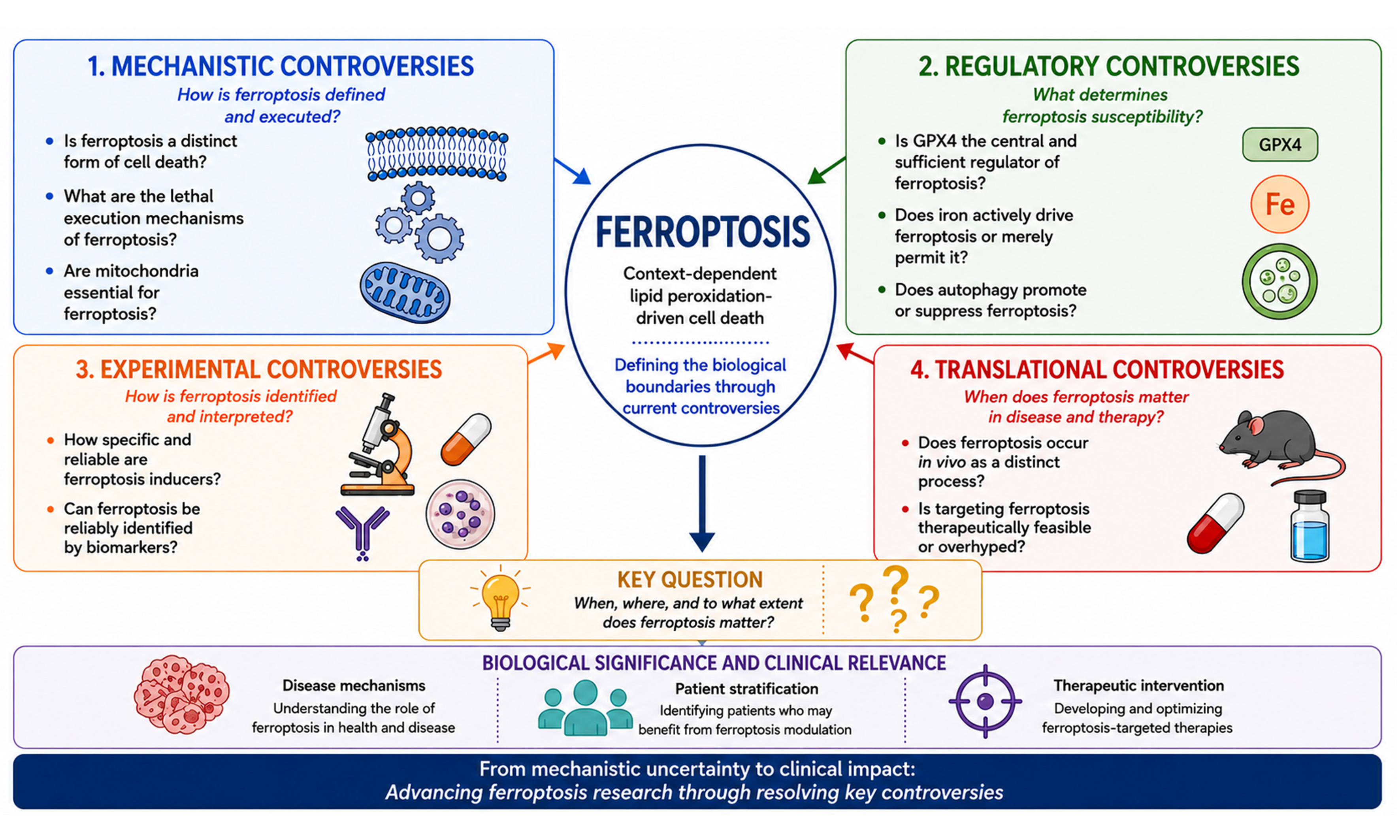
Ferroptosis is a form of regulated cell death that links iron metabolism, lipid peroxidation, and cellular stress responses. Despite substantial progress, several aspects of its definition, mechanisms, and biological significance remain under active investigation. In this review, we discuss ten key questions that continue to shape the field, covering mechanisms, regulatory networks, experimental approaches, and translational applications. These questions include whether ferroptosis represents a distinct cell death modality or a consequence of oxidative damage, the nature of its lethal execution mechanisms, and the context-dependent contributions of mitochondria, iron metabolism, and parallel antioxidant systems. We also discuss ongoing challenges related to the specificity of pharmacological tools and the lack of definitive biomarkers, which complicate the interpretation of ferroptosis, particularly in vivo. In addition, we consider the therapeutic potential of targeting ferroptosis in cancer and degenerative diseases, as well as the limitations associated with current approaches. By organizing these topics into four interconnected domains (mechanistic, regulatory, experimental, and translational), we provide a framework for understanding current challenges and unresolved issues in the field. Continued progress will likely benefit from improved biomarkers, rigorous experimental design, careful consideration of biological context, and the integration of systems-level approaches to facilitate therapeutic translation.
Keywords: Biomarkers, ferroptosis, iron metabolism, oxidative cell death, translational challenges
Key Points
• Ten unresolved controversies redefine the conceptual boundaries of ferroptosis.
• Mechanistic, regulatory, experimental, and translational challenges are systematically reviewed.
• Current evidence highlights context-dependent regulation of ferroptosis across biological systems.
• Limitations of biomarkers and experimental models complicate ferroptosis interpretation.
• Future priorities are proposed to improve mechanistic understanding and clinical translation.
INTRODUCTION
The maintenance of tissue homeostasis depends on a balance between cell survival and cell death. Beyond serving as a consequence of cellular injury, regulated cell death is an essential biological process that shapes development, removes damaged cells, and enables adaptation to environmental stress[1-4].
Over the past two decades, multiple forms of regulated cell death have been identified and characterized on the basis of their underlying molecular mechanisms and biological functions[5-7]. Among them, ferroptosis is distinguished by iron-dependent lipid peroxidation and the accumulation of oxidative damage in cellular membranes[8-10]. The discovery of ferroptosis has broadened our understanding of cell death regulation and revealed new links between metabolism, redox homeostasis, and disease[10-13]. Despite rapid progress, important questions remain regarding how ferroptosis should be defined, what molecular events ultimately drive cell death, and how these processes vary across biological contexts.
Since the term ferroptosis was introduced in 2012[14], interest in this field has grown rapidly across a wide range of experimental systems and disease settings[10-13]. However, the accumulation of new findings has not always led to consensus. Instead, differing interpretations and experimental limitations have continued to shape discussions in the field. In this review, we discuss ten unresolved questions related to ferroptosis and organize them into four broad areas: mechanisms, regulation, experimental models, and in vivo relevance [Table 1]. By bringing these issues together, we hope to facilitate discussion of areas of agreement and disagreement, draw attention to key knowledge gaps, and provide context for interpreting current challenges in the field.
Summary of the ten controversies guiding the next phase of ferroptosis research
| Controversy | Central question | Competing viewpoints | Key evidence | Potential resolution |
| 1 | Is ferroptosis a distinct form of cell death or part of a broader oxidative death continuum? | Unique regulated cell death pathway vs. oxidative death continuum | Distinct genetic/pharmacological regulation vs. overlap with oxytosis[120,238-242] | Adopt multi-parameter criteria integrating genetic, biochemical, and functional evidence |
| 2 | What is the ultimate executioner of ferroptosis? | Specific oxidized phospholipids vs. cumulative membrane damage | Oxidized phospholipid species vs. widespread membrane dysfunction[82,243-247] | Combine quantitative lipidomics with functional validation |
| 3 | Are mitochondria essential for ferroptosis? | Essential contributors vs. dispensable for core execution | Mitochondrial ROS vs. mitochondrial-deficient models[14,95,150,248-250] | Define stimulus-specific mitochondrial contributions |
| 4 | Is GPX4 the central and sufficient regulator? | Master regulator vs. multiple antioxidant systems | GPX4 knockout lethality vs. AIFM2/DHODH pathways[19,27,31,32,34,155,156,171,251-254] | Establish hierarchy of defense networks |
| 5 | Does iron actively drive ferroptosis or merely permit it? | Driver vs. permissive amplifier | Iron chelator protection vs. lipid composition effects[29,46,68,72,149,166,211,255] | Map iron flux, speciation, and distribution |
| 6 | Does autophagy promote or suppress ferroptosis? | Promoter vs. suppressor | Ferritinophagy vs. protective mitophagy[61,100,256-265] | Dissect selective autophagy pathways |
| 7 | Are ferroptosis inducers truly specific? | Reliable probes vs. off-target effects | Erastin/RSL3 utility vs. mixed death responses[196-198,266-268] | Develop more selective probes |
| 8 | Do we have reliable and specific biomarkers? | Current markers sufficient vs. non-specific | Lipid peroxidation assays vs. overlap with oxidative injury[269-272] | Integrate lipidomics and functional assays |
| 9 | Does ferroptosis occur in vivo as a distinct process? | Distinct in vivo pathway vs. indirect evidence | Animal studies vs. lack of definitive biomarkers[273-276] | Develop in vivo reporters and refined models |
| 10 | Is targeting ferroptosis therapeutically feasible or overhyped? | Therapeutic opportunity vs. translational barriers | Preclinical efficacy vs. toxicity/specificity concerns[83,153,226-232,235,237,277] | Use biomarker-guided, context-specific strategies |
CONCEPTUAL FRAMEWORK OF FERROPTOSIS
Ferroptosis can be considered as a disruption of cellular membrane homeostasis caused by the accumulation of phospholipid peroxidation[15-18]. Unlike apoptosis and necroptosis, ferroptosis has not yet been linked to a universally accepted dedicated execution protein[14]. Instead, ferroptosis is widely believed to involve the accumulation of lipid peroxidation beyond the capacity of cellular systems that prevent or repair oxidative membrane damage, although the molecular events linking lipid damage to cell death remain incompletely understood [Figure 1]. However, determining when these features are sufficient to attribute a biological phenotype to ferroptosis remains an important challenge in the field [Box 1].
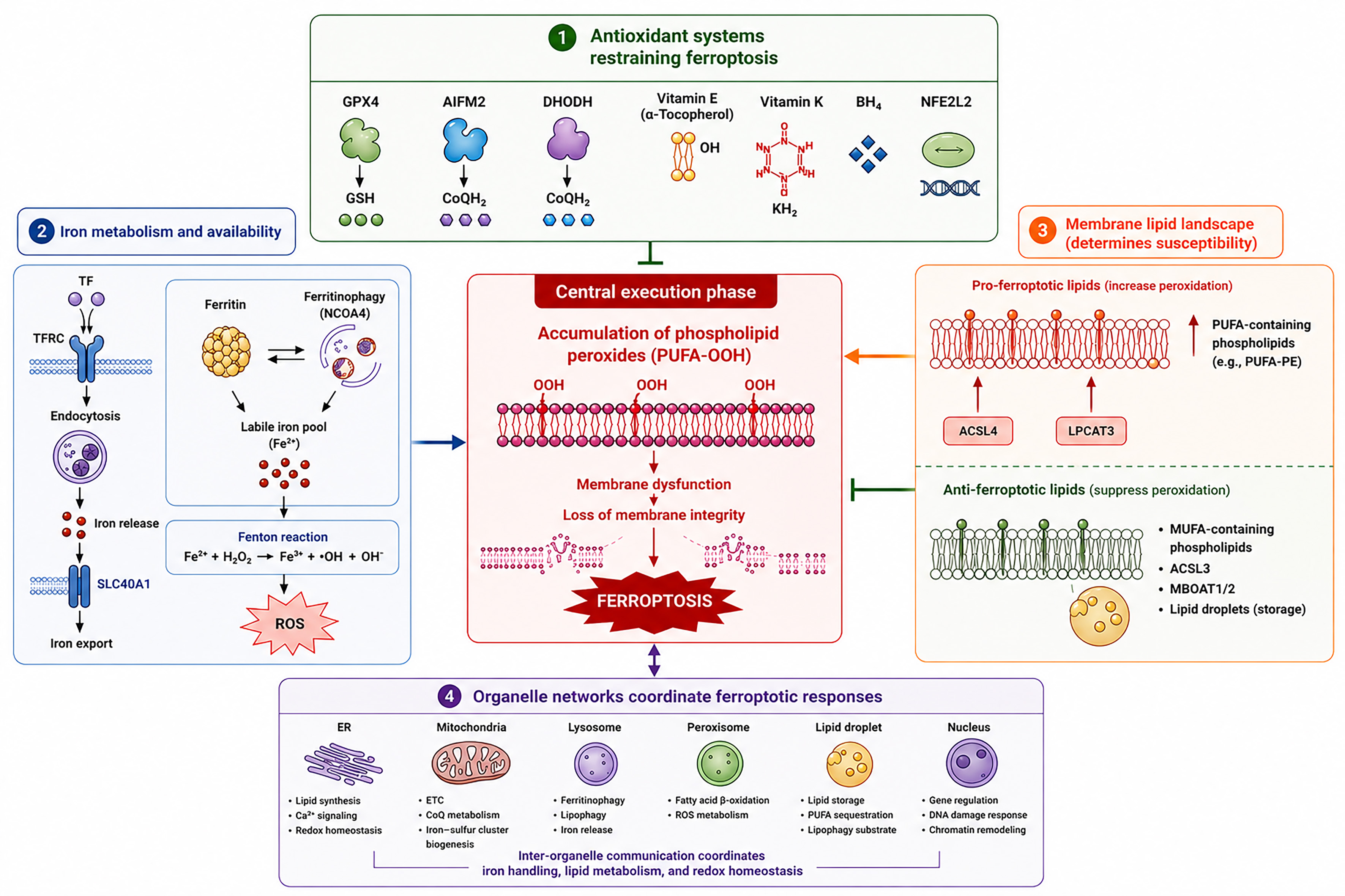
Figure 1. Conceptual framework of ferroptosis.
Ferroptosis is characterized by the accumulation of phospholipid peroxides beyond the capacity of cellular antioxidant and repair systems. Cellular susceptibility is determined by the interplay among four major regulatory modules: antioxidant defense systems, iron metabolism, membrane lipid composition, and organelle networks. Antioxidant pathways restrain phospholipid peroxidation, whereas iron availability and oxidation-prone membrane lipids promote oxidative membrane damage. Inter-organelle communication coordinates these processes through the regulation of iron handling, lipid metabolism, and redox homeostasis. Excessive phospholipid peroxidation ultimately leads to membrane dysfunction, loss of membrane integrity, and ferroptotic cell death. ACSL3: Acyl-CoA synthetase long-chain family member 3; ACSL4: acyl-CoA synthetase long-chain family member 4; AIFM2: apoptosis-inducing factor family member 2; BH4: tetrahydrobiopterin; CoQH2: reduced coenzyme Q (ubiquinol); DHODH: dihydroorotate dehydrogenase; DNA: deoxyribonucleic acid; ER: endoplasmic reticulum; ETC: electron transport chain; GPX4: glutathione peroxidase 4; GSH: glutathione; LPCAT3: lysophosphatidylcholine acyltransferase 3; MBOAT1/2: membrane-bound O-acyltransferase domain-containing 1/2; MUFA: monounsaturated fatty acid; NCOA4: nuclear receptor coactivator 4; NFE2L2: NFE2 like BZIP transcription factor 2; PUFA: polyunsaturated fatty acid; PUFA-OOH: polyunsaturated fatty acid hydroperoxide; PUFA-PE: polyunsaturated fatty acid-containing phosphatidylethanolamine; ROS: reactive oxygen species; SLC40A1: solute carrier family 40 member 1; TF: transferrin; TFRC: transferrin receptor.
Antioxidant systems restraining ferroptosis
A characteristic feature of ferroptosis is its sensitivity to pathways that detoxify phospholipid hydroperoxides before they accumulate. Among these, the glutathione peroxidase 4 (GPX4)-centered pathway remains the best-characterized defense mechanism. GPX4 utilizes glutathione (GSH) to reduce phospholipid hydroperoxides to lipid alcohols, thereby limiting the propagation of membrane oxidative damage[19]. Maintenance of this pathway depends on intracellular cysteine availability, which is largely supported by system xc--mediated cystine uptake[14]. Following import, cystine is reduced to cysteine, a rate-limiting precursor for GSH synthesis[20]. Disruption of cystine transport, depletion of GSH, or inhibition of GPX4 can therefore increase susceptibility to ferroptosis[19,21-23].
GPX4 exists as three isoforms that differ in their subcellular localization. Among these, the cytosolic isoform is generally considered the principal mediator of ferroptosis suppression, although the contributions of the mitochondrial and nuclear isoforms remain less well understood[19,24,25]. GPX4 is a selenoprotein whose activity depends on the incorporation of selenocysteine[26]. Recent studies have suggested that peroxiredoxin 6 (PRDX6) may contribute to intracellular selenium trafficking required for selenoprotein biosynthesis, including GPX4, and may also facilitate the membrane localization of GPX4[27-30]. Through these mechanisms, PRDX6 has been proposed to support GPX4-mediated detoxification of phospholipid hydroperoxides and the maintenance of membrane homeostasis during ferroptosis[27-30].
However, resistance to ferroptosis is not determined by GPX4 alone. Cells possess multiple antioxidant systems that can compensate when individual protective pathways are impaired. A well-characterized GPX4-independent defense mechanism is mediated by apoptosis-inducing factor (AIF) family member 2 [AIFM2, also known as ferroptosis suppressor protein 1 (FSP1)], which suppresses lipid peroxidation by reducing coenzyme Q (CoQ) to its antioxidant form, CoQH2, at cellular membranes[31,32]. Recent studies further suggest that AIFM2 may also contribute to ferroptosis resistance through the reduction of vitamin K[33,34]. Additional radical-trapping molecules, including vitamin E[14], reduced tetrahydrobiopterin[35,36], vitamin K[33,37], hydropersulfides[38,39], and serotonin-derived antioxidants[40], further contribute to limiting lipid radical propagation. The presence of these overlapping defense mechanisms may help explain why inhibition of a single pathway is often insufficient to induce ferroptosis.
At a broader regulatory level, both GPX4-dependent and GPX4-independent defense pathways are influenced by NFE2 like BZIP transcription factor 2 [NFE2L2, also known as nuclear factor erythroid 2-related factor 2 (NRF2)], a transcription factor that coordinates antioxidant and detoxification responses[41]. Under basal conditions, NFE2L2 is negatively regulated by Kelch-like ECH-associated protein 1 (KEAP1), which promotes its ubiquitination and proteasomal degradation. Oxidative or electrophilic stress disrupts KEAP1-mediated repression, allowing NFE2L2 to accumulate and induce the expression of genes involved in redox homeostasis, iron metabolism, GSH synthesis, and ferroptosis resistance[42-49]. Through the coordinated regulation of these pathways, NFE2L2 can influence cellular susceptibility to ferroptosis.
Iron availability as a catalyst of ferroptotic damage
Iron is an essential cofactor for numerous biological processes, including oxygen transport, mitochondrial metabolism, and DNA synthesis. In the context of ferroptosis, however, its importance derives from the availability of redox-active iron capable of promoting reactive oxygen species (ROS) generation and lipid peroxidation[14]. Cellular susceptibility therefore appears to correlate more closely with the size of the labile iron pool than with total iron content.
Cellular iron availability is regulated through a network of uptake, storage, utilization, and export pathways. A central regulator of these processes is the iron regulatory protein (IRP)-iron responsive element (IRE) system, which coordinates the expression of genes involved in iron uptake, storage, and export in response to intracellular iron status[50-52]. Changes in any of these processes can alter the size of the labile iron pool and consequently affect ferroptosis sensitivity[53,54]. Iron imported through transferrin receptor (TFRC)-mediated uptake can be mobilized for metabolic needs or stored in ferritin complexes[55-63], whereas excess iron may be exported through solute carrier family 40 member 1 [SLC40A1, also known as ferroportin (FPN)][64-68]. The balance among these pathways is dynamic and can vary across cell types and physiological conditions.
In addition to the overall size of the labile iron pool, the intracellular distribution of iron may also influence ferroptosis susceptibility. Mitochondria require iron for metabolic processes, including iron-sulfur cluster and heme biosynthesis, and can contribute to cellular ROS production. Alterations in mitochondrial iron homeostasis have therefore been proposed to modulate ferroptotic responses in some settings[69,70]. Lysosomes also contribute to intracellular iron homeostasis by regulating iron mobilization and recycling. Through ferritin degradation and related pathways, lysosomes can increase the availability of redox-active iron and thereby influence ferroptosis sensitivity[61,71,72]. Nevertheless, the relative contributions of mitochondrial and lysosomal iron metabolism to ferroptosis remain incompletely understood and may vary across biological contexts.
Membrane lipid composition determines ferroptotic susceptibility
The availability of oxidizable phospholipids is a critical determinant of ferroptosis susceptibility. Polyunsaturated fatty acid (PUFA)-containing phospholipids constitute the primary substrates for lipid peroxidation and are therefore closely linked to ferroptotic sensitivity[73-75]. Enzymes such as acyl-CoA synthetase long-chain family member 4 (ACSL4) and lysophosphatidylcholine acyltransferase 3 (LPCAT3) facilitate the incorporation of PUFAs into membrane phospholipids, generating lipid species that are particularly susceptible to oxidative modification[76-78]. In addition, cytochrome P450 oxidoreductase (POR), a nicotinamide adenine dinucleotide phosphate (NADPH)-dependent oxidoreductase, is implicated in the propagation of phospholipid peroxidation under certain conditions[17,79]. Consistent with these roles, genetic or pharmacological perturbation of lipid remodeling pathways can substantially alter cellular susceptibility to ferroptosis.
By contrast, pathways that enrich membranes with monounsaturated or saturated fatty acids generally confer protection[80-82]. Monounsaturated fatty acids (MUFAs) can compete with PUFAs for incorporation into membrane phospholipids, thereby reducing the abundance of oxidation-prone lipid species. These lipid-remodeling processes alter membrane composition in ways that reduce susceptibility to radical-mediated oxidation[83-85].
Ferroptosis sensitivity is influenced not only by the abundance of PUFAs, but also by the organization and trafficking of lipids within cells. Peroxisome-derived ether phospholipids increase ferroptosis susceptibility in some settings by providing additional substrates for lipid peroxidation, although their contribution appears to vary among cell types[86-88]. Lipid droplets may also influence ferroptosis by sequestering PUFAs and regulating their availability for membrane phospholipid synthesis[89-92].
Together, these findings suggest that ferroptosis susceptibility is shaped by the broader cellular lipid landscape, including lipid composition, distribution, and remodeling. Variability in these processes may contribute to the context-dependent nature of ferroptosis observed across different cell types and physiological conditions.
Organelle networks coordinate ferroptotic responses
Ferroptosis emerges from the integration of multiple metabolic processes that are distributed across different cellular organelles rather than being controlled by a single subcellular compartment. Mitochondria contribute to energy metabolism, redox homeostasis, and iron utilization[84,93-97]; lysosomes regulate iron recycling and nutrient turnover through degradative pathways[61,72,98-100]; the endoplasmic reticulum (ER) serves as a major site of lipid synthesis and membrane remodeling[16,101-104]; peroxisomes participate in ether phospholipid metabolism and fatty acid oxidation; and lipid droplets function as dynamic reservoirs that store and mobilize neutral lipids[86,89-92]. The Golgi apparatus contributes to membrane trafficking, lipid transport, and pH homeostasis[105,106], whereas the nucleus coordinates transcriptional programs that regulate cellular metabolism, stress responses, and ferroptosis-associated gene expression[107,108].
An important feature of this organization is the extensive communication between organelles. Iron, lipids, metabolites, and redox signals are continuously exchanged through interconnected metabolic networks, allowing changes in one compartment to influence ferroptosis-related processes in another[9]. Consequently, ferroptosis susceptibility is shaped not only by the activity of individual pathways but also by the coordination of organelle functions at the cellular level.
This systems-level organization may help explain the context-dependent nature of ferroptosis. Differences in metabolic state, organelle activity, or inter-organelle communication can alter the relative importance of specific ferroptosis regulators across cell types and experimental conditions. As a result, mechanisms identified as critical in one setting may play a more limited role in another.
Execution phase: from lipid peroxidation to membrane failure
The later stages of ferroptosis are associated with the accumulation of lipid peroxidation products within cellular membranes. Unlike apoptosis and necroptosis, which involve defined protein effectors that execute cell death, ferroptosis has not yet been linked to a universally accepted execution mechanism. Nevertheless, progressive lipid damage is widely considered a central event preceding loss of membrane integrity[109-112].
Current models propose that extensive phospholipid peroxidation alters membrane organization, biophysical properties, and cellular homeostasis, ultimately contributing to membrane dysfunction and cell death. However, the molecular events that connect lipid damage to terminal membrane failure remain incompletely understood. Whether membrane rupture results directly from the accumulation of oxidized phospholipids or requires additional downstream processes remains an area of active investigation.
Membrane failure is not determined solely by the extent of oxidative damage. Cells possess repair mechanisms that can counteract membrane injury, including endosomal sorting complex required for transport III (ESCRT-III)-mediated repair pathways[113,114]. The balance between ongoing membrane damage and repair capacity may therefore influence the timing and outcome of ferroptotic cell death.
Defining how oxidized membranes ultimately lose integrity remains an important challenge for the field. Addressing this question may help clarify whether ferroptosis is executed primarily through biophysical membrane damage, specialized molecular mechanisms, or a combination of both.
TEN CONTROVERSIES DEFINING THE BOUNDARIES OF FERROPTOSIS
Although ferroptosis is now widely recognized as a form of regulated cell death, many fundamental questions remain unresolved. As the field has expanded, increasing mechanistic complexity and diverse experimental observations have often generated new debates rather than consensus. These controversies extend beyond individual molecular pathways and reflect broader challenges in defining the conceptual boundaries of ferroptosis. To facilitate discussion, the ten controversies outlined below are organized into four interconnected domains: mechanistic definition, regulatory control, experimental interpretation, and translational relevance [Figure 2]. Together, they address how ferroptosis should be defined, what determines ferroptotic susceptibility and execution, how it can be rigorously identified, and when it contributes meaningfully to physiology, pathology, and therapeutic intervention.
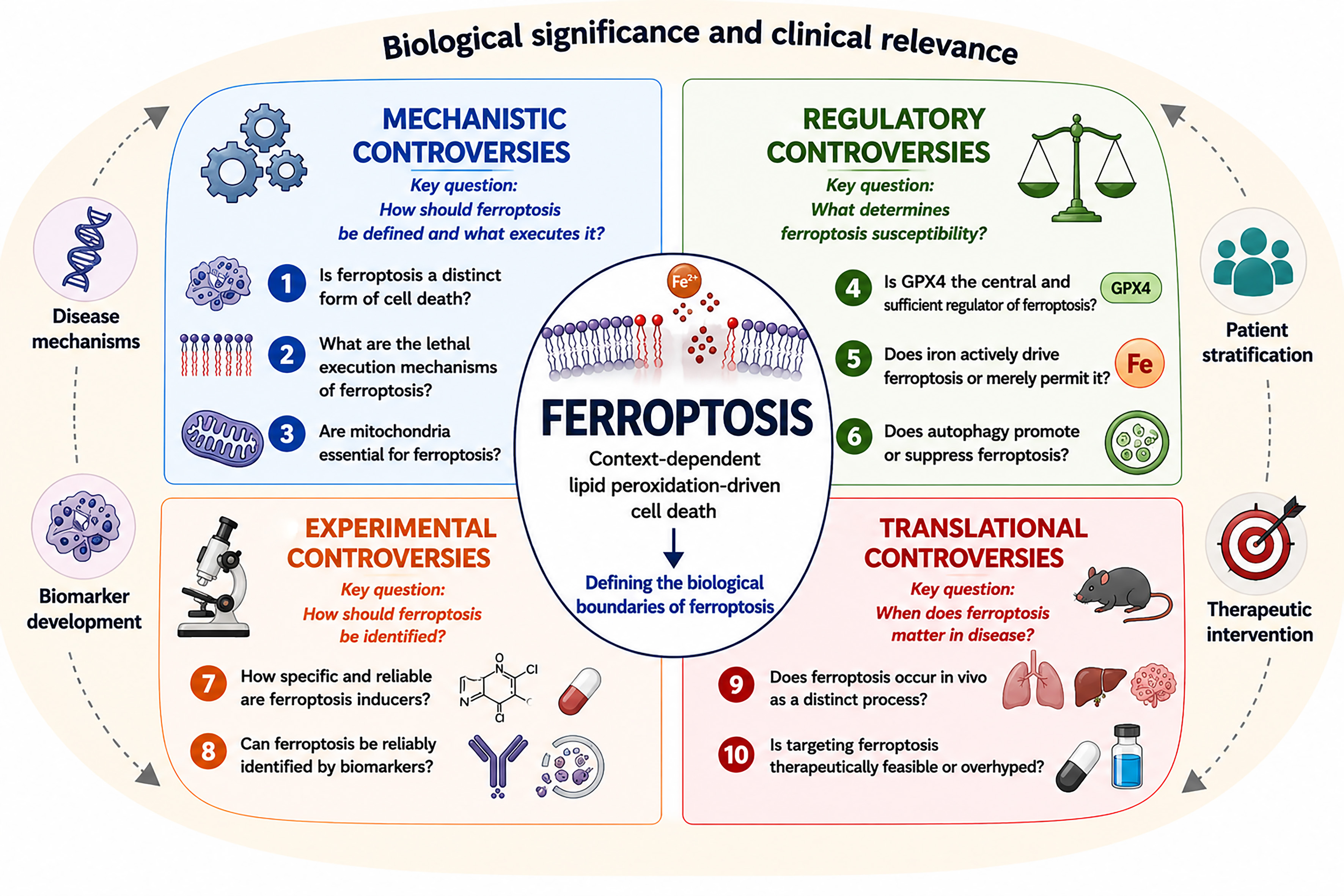
Figure 2. Current controversies defining the boundaries of ferroptosis.
The ten controversies discussed in this review are grouped into four interconnected domains: mechanistic definition, regulatory control, experimental interpretation, and translational relevance. These debates encompass questions regarding the identity and execution of ferroptosis, the pathways that govern ferroptosis susceptibility, the criteria used for its experimental attribution, and its significance in disease and therapeutic intervention. GPX4: Glutathione peroxidase 4.
Mechanistic controversies
Controversy 1: Is ferroptosis a distinct form of cell death or part of a broader oxidative death continuum?
Over the past decade, the Nomenclature Committee on Cell Death (NCCD) has established guidelines for defining and classifying cell death based on morphological, biochemical, and functional criteria[115]. Within these guidelines, cell death is generally categorized into accidental cell death and regulated cell death. Ferroptosis has been widely recognized as a form of regulated cell death characterized by iron-dependent lipid peroxidation and the failure of specific antioxidant defense systems, most notably GPX4. Genetic ablation of Gpx4 or disruption of cystine uptake via system xc- is sufficient to induce cell death with hallmark lipid peroxidation signatures, whereas this process can be selectively suppressed by lipophilic antioxidants (e.g., ferrostatin-1 and liproxstatin-1) or iron chelators (e.g., deferoxamine)[14,116-118]. In addition, ferroptosis displays morphological and biochemical features that are distinct from apoptosis and necroptosis, and its execution is not prevented by inhibitors of these pathways, such as Z-VAD-FMK or necrosulfonamide[14]. Together, these findings provide a framework for understanding ferroptosis as a regulated form of cell death with identifiable molecular features and pharmacological vulnerabilities.
An alternative perspective, however, argues that ferroptosis may not represent a fundamentally distinct entity, but rather a manifestation of oxidative stress-induced cell death within a broader continuum[119]. Oxidative stress is not a singular signal, but a spectrum of molecular damage and signaling events whose outcomes depend on intensity, localization, and cellular context. Thus, cells may engage different death programs in response to similar oxidative insults[119]. Ferroptosis shares key features with earlier-described oxidative damage processes such as oxytosis, first identified in neurons in response to glutamate-induced inhibition of the cystine-glutamate antiporter[120-122]. Both processes involve depletion of GSH, inactivation of GPX4, and the accumulation of iron-dependent lipid peroxidation. Even earlier studies by Harry Eagle in the mid-20th century revealed that nutrient deprivation, particularly of cysteine, can induce cell death in mouse fibroblasts and the human HeLa cancer cell line[123], suggesting that core elements of ferroptosis-like processes have long been recognized.
Lipid peroxidation, ROS accumulation, and membrane damage are not unique to ferroptosis and can also be observed in other forms of cellular injury, including apoptosis[124-128], pyroptosis[129-132] and necroptosis[133]. This overlap has led to the suggestion that some features commonly associated with ferroptosis may reflect particular metabolic conditions, such as increased polyunsaturated lipid content or altered iron metabolism, rather than a completely distinct execution mechanism. Supporting this possibility, several studies have reported concurrent activation of ferroptotic and non-ferroptotic death pathways under conditions of severe stress, while the absence of definitive biomarkers continues to complicate the identification of ferroptosis in vivo[134-136]. Recent work further indicates that ferroptosis propagation is often accompanied by heterogeneous cell death profiles that include features of apoptosis and necrosis[137]. In tissue injury settings, ferroptosis may therefore occur alongside other forms of cell death rather than as an isolated process, making it difficult to distinguish its specific contribution to pathology.
The persistence of this debate reflects a broader challenge in cell death research: biological processes rarely conform to rigid categorical boundaries. Rather than asking whether ferroptosis is entirely distinct from other forms of oxidative cell death, it may be more useful to identify the contexts in which its defining molecular features become biologically consequential. Such efforts should help refine the conceptual boundaries of ferroptosis while clarifying its relevance to human disease.
Controversy 2: What is the ultimate executioner of ferroptosis?
Ferroptosis culminates in the loss of plasma membrane integrity, yet the molecular events responsible for this transition remain incompletely defined. Because the plasma membrane is highly enriched in phospholipids, extensive lipid peroxidation has long been proposed to compromise membrane structure and function. Consistent with this view, ferroptosis ultimately results in membrane rupture and the release of intracellular contents, including damage-associated molecular patterns (DAMPs), which may influence the immunological consequences of cell death[138]. Despite this lytic outcome, ferroptosis differs mechanistically from other forms of regulated necrosis. In necroptosis and pyroptosis, membrane disruption is mediated by dedicated pore-forming effectors, most notably mixed lineage kinase domain-like pseudokinase (MLKL) and members of the gasdermin family[8,139]. By contrast, no universally accepted executioner protein has yet been identified for ferroptosis, raising the question of how lipid peroxidation is translated into terminal membrane failure.
A commonly proposed mechanism is that ferroptosis is driven by the accumulation of oxidized lipid species that progressively disrupt membrane integrity [Table 2]. In this model, PUFA-containing phospholipids, particularly phosphatidylethanolamines (PUFA-PEs), are selectively oxidized through enzymatic and non-enzymatic reactions to generate cytotoxic lipid peroxides[140]. Consistent with this model, genetic or pharmacological interventions that limit the generation of peroxidation-prone phospholipids reduce ferroptosis sensitivity, whereas inhibition of lipid peroxide detoxification promotes cell death[76-78]. Detoxification of lipid peroxides by GPX4 or radical-trapping antioxidants, such as endogenous vitamin E, 7-dehydrocholesterol, or synthetic agents such as ferrostatin-1, suppresses ferroptotic cell death, supporting the notion that lipid peroxidation represents a proximal execution mechanism[19,26,141-143]. In contrast, ACSL3-dependent incorporation of MUFAs into membrane phospholipids reduces susceptibility to peroxidation and confers resistance to ferroptosis[80,92]. Membrane-bound O-acyltransferase domain-containing proteins 1 and 2 (MBOAT1/2) are identified as ferroptosis suppressors that remodel phospholipid composition in a MUFA-dependent manner, although their functions are distinct from canonical MUFA incorporation pathways[144]. Although these studies support a role for specific oxidized phospholipids in ferroptotic execution, whether any individual lipid species is necessary or sufficient to drive cell death remains uncertain.
Major regulatory pathways controlling ferroptosis and their context-dependent functions
| Regulatory pathway | Representative components | Proposed anti-ferroptotic function | Proposed pro-ferroptotic function | Context-dependent considerations |
| GPX4-GSH pathway | System xc-, cysteine, GSH, GPX4[19] | Detoxifies phospholipid hydroperoxides and preserves membrane integrity | Loss of GPX4 permits toxic lipid peroxide accumulation | Primary defense pathway but may be compensated by alternative systems |
| AIFM2-CoQ10 pathway | AIFM2/FSP1, CoQ10, NAD(P)H[31,32] | Traps lipid radicals independently of GPX4 | Loss of AIFM2 increases ferroptosis sensitivity | Importance varies among cell types and metabolic states |
| DHODH pathway | DHODH, mitochondrial CoQ[278] | Limits mitochondrial lipid peroxidation | DHODH inhibition sensitizes cells to ferroptosis | Particularly important in mitochondria-rich cells |
| GCH1-BH4 pathway | GCH1, BH4[35,36] | Protects membrane lipids and supports antioxidant defense | BH4 depletion enhances oxidative damage | Contribution varies across tissues and disease settings |
| Iron metabolism | TFRC, ferritin, NCOA4, SLC40A1/ferroportin[61,64,100] | Iron sequestration reduces oxidative stress | Iron uptake and ferritinophagy promote ferroptosis | Driver versus amplifier role remains debated |
| Lipid metabolism | ACSL4, LPCAT3, ALOXs[76-78] | Reduced PUFA incorporation decreases susceptibility | PUFA phospholipids provide oxidation substrates | Major determinant of ferroptosis sensitivity |
| MUFA remodeling pathway | ACSL3, MUFAs, MBOAT1/2[80,92,144] | MUFA incorporation suppresses ferroptosis | Loss of remodeling increases susceptibility | Depends on cellular lipid remodeling capacity |
| Autophagy-lysosome pathway | NCOA4, ATGs, lysosomes[61,71,72,100] | Mitophagy may reduce oxidative stress | Ferritinophagy and lipophagy promote ferroptosis | Can suppress or promote ferroptosis depending on context |
| NFE2L2 signaling | NFE2L2, KEAP1[42-49] | Coordinates antioxidant defense and iron homeostasis | Excessive HMOX1 may increase iron availability | Effects vary with redox status and tissue context |
| Mitochondrial metabolism | TCA cycle, ETC complexes, β-oxidation[84,97] | Supports redox homeostasis | ROS generation can enhance ferroptosis | Depends on initiating stimulus |
An alternative interpretation is that no single lipid species acts as the definitive executioner of ferroptosis. Instead, cell death may result from the cumulative disruption of membrane homeostasis caused by widespread lipid peroxidation. In this model, the overall burden of oxidative damage across cellular membranes progressively compromises membrane integrity, ultimately leading to membrane rupture[145]. Consistent with this model, oxidized lipids accumulate across multiple membrane compartments during ferroptosis, suggesting that overall membrane damage rather than a single lipid species may determine cell fate[82,86,146]. The identity and contribution of putative “lethal” lipid species also vary across cell types and experimental systems, indicating that ferroptotic execution may be context-specific.
Despite this distinction, emerging evidence suggests that ferroptosis may engage additional execution-related processes beyond lipid peroxidation. This possibility has attracted attention because several proteins traditionally associated with other lytic death pathways have been implicated in ferroptotic settings. For example, activation of gasdermin E (GSDME), a pore-forming protein associated with pyroptosis, has been reported to contribute to ferroptosis in specific in vivo contexts[147]. These findings indicate that ferroptosis may incorporate elements of other lytic pathways under certain conditions, although lipid peroxidation remains central to its definition.
The key question is not whether lipid peroxidation occurs during ferroptosis, but whether it is itself sufficient to execute cell death. Although oxidized phospholipids are a defining feature of ferroptotic cells, it remains unclear whether they directly drive membrane failure or act upstream of additional execution mechanisms. Resolving this distinction will be essential for determining whether ferroptosis is ultimately governed by discrete molecular effectors, cumulative membrane damage, or both.
Controversy 3: Are mitochondria essential for ferroptosis?
Like other forms of regulated cell death, the initiation and regulation of ferroptosis involve multiple organelles. Among these, the ER plays a key role in ferroptosis due to its function as a major site of lipid synthesis and remodeling[103,104]. However, the role of mitochondria in ferroptosis remains controversial. Numerous studies have implicated mitochondria in ferroptosis, particularly under conditions of metabolic stress. Through their roles in the tricarboxylic acid cycle, electron transport, iron-sulfur cluster biogenesis, fatty acid β-oxidation, and ROS production, mitochondria can influence the metabolic and redox conditions that favor ferroptosis. Consistent with this view, cystine deprivation enhances mitochondrial metabolism and ROS generation, thereby increasing ferroptosis susceptibility[93]. Characteristic mitochondrial morphological changes, including condensed membrane densities and reduced cristae, are also frequently observed during ferroptotic cell death[14].
Recent studies further extend this model by identifying specific mitochondrial-lipid interactions that modulate ferroptosis. For example, phospholipids containing two polyunsaturated fatty acyl chains (PL-PUFA2s), including PC-PUFA2s, can interact with mitochondrial electron transport chain complex I and promote mitochondrial ROS production, thereby facilitating ferroptosis[84]. Mitochondrial cellular communication network factor 1 (CCN1) also drives ferroptosis through the promotion of fatty acid β-oxidation and mitochondrial ROS production[97]. Conversely, cytochrome c (CYCS) released into the cytosol may inhibit ferroptosis by interacting with inositol polyphosphate-4-phosphatase type I A (INPP4A) and modulating phosphatidylinositol metabolism[148]. Thus, mitochondria can shape ferroptotic sensitivity through metabolic and signaling pathways. These findings also suggest that inter-organelle communication, particularly between mitochondria and other subcellular compartments such as the ER and lysosomes, may further regulate susceptibility to ferroptosis[16,149].
In contrast, other studies indicate that mitochondria are not essential for ferroptosis, challenging their role as core components of the execution machinery. Cells lacking functional mitochondria, including mitochondrial DNA-deficient (ρ0) cells, remain capable of undergoing ferroptosis, particularly when GPX4 is directly inhibited[14]. However, ρ⁰ cells exhibit broad metabolic and redox alterations beyond the loss of mitochondrial DNA[150], which may influence ferroptosis sensitivity and should be taken into account when interpreting these findings. Similarly, ferroptosis induced by GPX4 inhibitors, such as RSL3, proceeds independently of mitochondrial metabolism, relying instead on lipid peroxidation at cellular membranes[93,151]. In these contexts, iron-dependent lipid peroxide accumulation and cell death can occur without detectable contributions from mitochondrial ROS or bioenergetics.
Overall, the key question may not be whether mitochondria participate in ferroptosis, but whether they are required for its execution. Current evidence indicates that mitochondrial activity can strongly influence ferroptosis under specific metabolic conditions, yet ferroptotic death can also occur in their absence. These findings suggest that mitochondria function as important modulators of ferroptosis in some settings rather than as universally required components of the ferroptotic machinery.
Regulatory controversies
Controversy 4: Is GPX4 the central and sufficient regulator of ferroptosis?
GPX4 is widely regarded as the canonical suppressor of ferroptosis because of its ability to reduce phospholipid hydroperoxides within cellular membranes and limit the accumulation of lipid peroxidation products[152]. Consistent with this role, genetic deletion of Gpx4 induces ferroptotic cell death in multiple tissues, whereas pharmacological inhibition of GPX4 robustly triggers ferroptosis across diverse experimental systems[116,153]. Whole-body Gpx4 knockout is embryonically lethal in mice, underscoring the importance of GPX4 for maintaining cellular viability and redox homeostasis[154].
Despite these observations, whether GPX4 is universally required or sufficient to determine ferroptosis sensitivity remains debated. As discussed above, AIFM2 represents one of the best-characterized GPX4-independent ferroptosis defense pathways[155,156]. Upon myristoylation-dependent translocation to the plasma membrane, AIFM2 is thought to reinforce membrane-associated antioxidant capacity and limit lipid peroxidation[31,32]. The discovery of this pathway demonstrated that ferroptosis resistance can, under certain conditions, be maintained independently of GPX4, indicating that multiple protective systems contribute to ferroptosis regulation[155,156].
4-hydroxynonenal (4-HNE) is a highly reactive α,β-unsaturated aldehyde generated during the peroxidation of ω-6 PUFAs, particularly arachidonic acid and linoleic acid. As one of the most extensively investigated secondary products of lipid peroxidation, it can directly induce ferroptosis[157]. Of note, 4-HNE-mediated adduction of AIFM2 can alter its function, promoting its translocation to the nucleus and shifting its activity from an NAD(P)H oxidoreductase toward a pro-apoptotic role[158], suggesting that excessive lipid peroxidation may redirect cell death programs from ferroptosis toward apoptosis. Conversely, cells with intact GPX4 can still exhibit ferroptotic sensitivity when oxidative stress exceeds the combined capacity of these defense pathways. In addition, other GPX family members (e.g., GPX1[159,160] and GPX7[161-163]) inhibit ferroptosis in a context-specific manner, suggesting that their functions may be partially complementary.
Genetic studies further illustrate the complexity of ferroptosis regulation. Although global Gpx4 deletion causes embryonic lethality, global Aifm2 knockout mice remain viable. In addition, tissue-specific Gpx4 deletion produces markedly different phenotypes across organs, causing spontaneous severe injury in some tissues, such as the kidney[116], whereas producing more limited or context-dependent effects in others, including the pancreas[164]. Such variability suggests that the relative contribution of GPX4 depends on cellular context, metabolic state, and the availability of alternative defense mechanisms.
Collectively, these findings indicate that GPX4 is a major regulator of ferroptosis but not necessarily its sole determinant. The central question is whether ferroptosis is organized around GPX4 as a dominant defense pathway or a broader network of partially overlapping protective mechanisms. Resolving this issue will likely depend on genetic strategies capable of defining when GPX4-dependent and GPX4-independent defenses function redundantly, cooperatively, or independently across different biological contexts.
Controversy 5: Does iron actively drive ferroptosis or merely permit it?
The requirement for iron is one of the most distinctive features of ferroptosis, yet its role in the process remains a matter of discussion. Whether iron functions as a direct mediator of cell death or primarily enables lipid peroxidation is not fully understood. A commonly proposed mechanism is that iron promotes ferroptosis through its participation in redox reactions that generate lipid-damaging radicals. The labile iron pool can support Fenton chemistry, whereby hydrogen peroxide is converted into highly reactive hydroxyl radicals capable of initiating and propagating lipid peroxidation[165]. Substantial genetic and pharmacological evidence supports this model: increasing intracellular iron levels enhances ferroptotic sensitivity, whereas iron chelators such as deferoxamine potently suppress ferroptotic cell death[14]. In addition, endogenous iron-binding molecules, including polyamines such as spermine, limit ferroptosis in certain tumors and tissue injury models[166]. Pathways that mobilize stored iron, such as ferritin degradation (ferritinophagy), further promote ferroptosis[61,100], reinforcing the idea that iron availability directly influences its execution. Although iron is distributed across multiple organelles, lysosomal iron may play a particularly important role in ferroptosis initiation, potentially by serving as a readily mobilizable redox-active pool[71,72]. Together, these findings position iron as a major catalytic contributor that can drive oxidative membrane damage.
In contrast, a complementary view proposes that iron is not necessarily the primary driver but rather a permissive factor that enables ferroptosis under specific metabolic conditions. From this perspective, lipid peroxidation can be initiated through enzymatic pathways, such as lipoxygenase-mediated oxidation of polyunsaturated phospholipids [e.g., arachidonate 15-lipoxygenase (ALOX15)-phosphatidylethanolamine binding protein 1 (PEBP1)-dependent phosphatidylethanolamine oxidation[167]], and subsequently propagated via non-enzymatic radical chain reactions[168]. Once initiated, these lipid peroxidation chains can proceed without continuous iron catalysis, relying instead on self-sustaining radical propagation within membrane lipids. The requirement for iron may therefore reflect its role in establishing or amplifying a pro-oxidant environment rather than serving as the central executor of cell death. Supporting this interpretation, modulation of lipid composition (e.g., PUFA availability) or antioxidant capacity can strongly influence ferroptotic sensitivity, in some cases overriding the effects of altered iron levels[19,84,169-172]. Moreover, ferroptotic susceptibility varies across cell types and conditions despite comparable intracellular iron content, highlighting the importance of additional regulatory factors.
Thus, whether iron is considered a driver or a permissive factor may depend on the biological scale being examined. Although iron provides the redox potential that underlies lipid oxidation, its effects are ultimately shaped by the metabolic and antioxidant networks in which it operates.
Controversy 6: Does autophagy promote or suppress ferroptosis?
Early studies suggested that ferroptosis may be independent of autophagy, based in part on observations that the lysosomal inhibitor chloroquine failed to block erastin-induced cell death[14]. However, this interpretation has certain limitations. Chloroquine disrupts lysosomal function and affects multiple pathways beyond autophagy, and its experimental use can be confounded by variability in stability and dosing[173]. Thus, these early findings do not definitively exclude a role for autophagy in ferroptosis.
Subsequent studies suggest that excessive autophagy and lysosomal activation can promote ferroptosis, at least in part, by facilitating iron mobilization and enhancing lipid peroxidation under various pathological conditions[174-181]. A central mechanism is ferritinophagy, a selective form of autophagy mediated by nuclear receptor coactivator 4 (NCOA4) that targets ferritin for lysosomal degradation, thereby increasing the labile iron pool[61,100]. Genetic inhibition of core autophagy components or NCOA4 suppresses ferroptosis in multiple models, whereas activation of autophagy can sensitize cells to ferroptotic stimuli[56,182-187]. Upstream regulatory pathways also contribute: for example, the E3 ligase membrane associated Ring-CH-type finger 7 (MARCHF7, also known as MARCH7) promotes proteasomal degradation of NCOA4, thereby limiting ferritinophagy and reducing ferroptosis[188]. Beyond iron metabolism, autophagic processes such as lipophagy may increase the availability of PUFAs, providing substrates for lipid peroxidation[189-191]. Collectively, these findings support a model in which autophagy acts upstream of ferroptosis by remodeling intracellular metabolism in a manner that favors oxidative membrane damage.
In contrast, an alternative perspective emphasizes the cytoprotective functions of autophagy, arguing that it can suppress ferroptosis by maintaining cellular homeostasis and limiting oxidative stress. Autophagy contributes to the clearance of damaged mitochondria (mitophagy) and oxidized macromolecules, thereby reducing sources of ROS and preventing the accumulation of toxic lipid peroxides[192-194]. GPX4 stabilizes BCL2 interacting protein 3 (BNIP3), thereby promoting mitophagy and reinforcing mitochondrial quality control[195]. These findings challenge a strictly pro-ferroptotic model and instead suggest that autophagy can buffer against ferroptotic damage under certain conditions.
Thus, the relationship between autophagy and ferroptosis may be better understood as a question of selectivity rather than directionality. Different autophagic pathways influence iron metabolism, lipid homeostasis, and organelle quality control in distinct ways, making it unlikely that autophagy exerts a uniformly pro- or anti-ferroptotic effect across all biological settings.
Experimental controversies
Controversy 7: Are ferroptosis inducers truly specific?
Canonical ferroptosis inducers are commonly used to investigate ferroptotic cell death and have provided important insights into its regulation and execution. Compounds such as erastin and RSL3 have been instrumental in defining the field by targeting key nodes of ferroptosis regulation: system xc--mediated cystine uptake and GPX4 activity, respectively[14,19]. These agents reproducibly induce iron-dependent lipid peroxidation and cell death that can often be rescued by ferroptosis inhibitors, including lipophilic radical-trapping antioxidants and iron chelators. Their consistent use across diverse models has helped establish operational criteria for ferroptosis and enabled the identification of many genetic regulators.
However, accumulating evidence raises concerns that these compounds are not fully pathway-specific. Erastin was originally identified as a mutant RAS-selective lethal compound and has been reported to target mitochondrial voltage-dependent anion channels (VDACs), including VDAC2 and VDAC3, in addition to inhibiting system xc-, suggesting that it can influence mitochondrial metabolism and broader cellular stress responses[196]. RSL3 and related GPX4 inhibitors are more direct tools for disabling lipid peroxide repair, but concerns remain regarding off-target effects, including reported activity toward other redox enzymes such as thioredoxin reductase 1 (TXNRD1)[197]. RSL3 can also trigger pyroptosis-associated features in cancer cells, including gasdermin cleavage and inflammatory cytokine release[198], indicating that ferroptotic stress may engage convergent cell death programs in some contexts. The use of relatively high concentrations of RSL3 (e.g., in the micromolar range) in some studies may induce mixed or non-canonical forms of cell death, thereby complicating mechanistic attribution.
Other commonly used ferroptosis inducers illustrate the same issue. Sorafenib can inhibit system xc- and induce ferroptosis in some settings[45,102,199], but it is also a multi-kinase inhibitor and can trigger non-ferroptotic death at higher concentrations, making mechanistic attribution difficult[200,201]. Sulfasalazine inhibits system xc-, but evidence for its ability to induce bona fide ferroptosis is context-dependent and less consistent[202,203]. FIN56 induces ferroptosis through at least two distinct mechanisms, namely GPX4 degradation and CoQ10 depletion downstream of squalene synthase activation[204]. In addition, FINO2 induces ferroptosis by indirectly impairing GPX4 activity while simultaneously oxidizing labile iron, thereby promoting lipid peroxidation[205]. These examples highlight the mechanistic complexity of ferroptosis induction and illustrate that even widely used ferroptosis inducers often engage multiple pathways rather than acting through a single, well-defined target.
A balanced interpretation is that current ferroptosis inducers are indispensable experimental tools, yet their specificity should not be presumed. This limitation extends beyond pharmacological agents to the biological contexts in which ferroptosis is studied. Widely used pathological models, such as ischemia-reperfusion injury[206,207], acute kidney injury[208-210], and neurodegeneration[211,212], engage complex and overlapping cell death programs, including apoptosis, necroptosis, and inflammatory responses. Consequently, ferroptotic features observed under these conditions may represent one component of a broader, integrated stress response rather than an isolated execution pathway.
Given these limitations, responses to canonical ferroptosis inducers should be interpreted with appropriate caution and not regarded as definitive evidence of ferroptosis in isolation. Rather, confidence in ferroptosis attribution increases when pharmacological observations are supported by complementary biochemical and genetic evidence. Continued refinement of experimental standards and chemical tools should improve the interpretation of ferroptosis studies and help distinguish ferroptosis-driven phenotypes from broader oxidative stress responses.
Controversy 8: Do we have reliable and specific biomarkers of ferroptosis?
A common assumption is that combining established biochemical and functional readouts is generally sufficient to identify ferroptosis. Lipid peroxidation assays, such as boron-dipyrromethene undecanoic acid (BODIPY-C11) oxidation, together with measurements of malondialdehyde (MDA) and 4-HNE, are widely used to detect oxidative damage to cellular membranes. In parallel, depletion or inactivation of GPX4, accumulation of labile iron, and sensitivity to ferroptosis inhibitors are often considered supportive evidence. When these features are observed together, many studies interpret them as indicative of ferroptotic cell death. This multi-parameter approach, particularly when combined with genetic manipulation and pharmacological rescue, has enabled reproducible identification of ferroptosis across diverse experimental systems.
In contrast, a growing body of work argues that none of these markers are truly specific to ferroptosis, raising concerns about overinterpretation. Lipid peroxidation is a general consequence of oxidative stress and occurs in multiple forms of cell death and tissue injury. Similarly, increases in MDA and 4-HNE reflect nonspecific membrane damage rather than a ferroptosis-defining signature. Even functional criteria, such as sensitivity to antioxidants or iron chelators, can overlap with other oxidative processes. Expression changes in genes involved in iron and lipid metabolism, such as ACSL4[76] and TFRC[213], or oxidation states of proteins such as peroxiredoxin 3 (PRDX3)[214], are frequently associated with ferroptosis but are not uniquely specific and may reflect broader metabolic remodeling.
In addition, ferroptosis is accompanied by the release of DAMPs, including high mobility group box 1 (HMGB1) and adenosine triphosphate (ATP)[111,215]; however, these signals are not unique to ferroptosis and are shared across multiple cell death modalities, limiting their specificity. Molecules such as decorin (DCN)[216] and GPX4[217] are proposed as potential ferroptosis-associated DAMPs, but their specificity, mode of release, and relevance under physiological or inflammatory conditions remain incompletely defined. The absence of a single, definitive biomarker makes it difficult to distinguish ferroptosis from related cell death modalities, particularly in vivo, where complex and overlapping pathways are engaged.
These limitations highlight a broader challenge: the search for a ferroptosis biomarker may ultimately be more complex than the identification of a single molecular signature. Because ferroptosis reflects the integration of iron metabolism, lipid peroxidation, antioxidant defenses, and cellular context, no individual marker is likely to capture all ferroptotic states. Greater diagnostic confidence may therefore depend on combinations of biochemical, genetic, and functional indicators rather than any single readout. Whether a universally applicable ferroptosis biomarker exists remains uncertain, but resolving this issue will be critical for defining the contribution of ferroptosis in physiological and disease settings.
Translational controversies
Controversy 9: Does ferroptosis occur in vivo as a distinct process?
Substantial evidence supports the occurrence of ferroptosis in vivo and suggests that it contributes to a range of physiological and pathological processes. In multiple disease models, including cancer[155,156,218], acute kidney injury[208,219,220], and neurodegenerative disorders[221,222], hallmarks associated with ferroptosis (such as iron accumulation, lipid peroxidation, altered ferroptosis-related gene expression, and responsiveness to ferroptosis-modulating interventions) have been reported. Genetic and pharmacological studies further indicate that manipulation of ferroptosis-related pathways can influence disease progression, supporting the biological relevance of ferroptosis beyond cultured cells.
Nevertheless, establishing ferroptosis in vivo remains considerably more challenging than demonstrating it in simplified experimental systems. Most evidence relies on combinations of indirect observations rather than direct visualization of ferroptotic cell death. Several hallmarks commonly used to infer ferroptosis are shared with other forms of tissue injury, complicating the interpretation of in vivo findings[119,223,224]. In addition, pharmacological interventions may exert broader antioxidant or metabolic effects that are not exclusively linked to ferroptosis inhibition. These limitations complicate the interpretation of in vivo studies and raise questions regarding the strength of evidence required to attribute pathological outcomes to ferroptosis.
An additional challenge arises from the complexity of tissue environments. Cell populations within the same tissue often differ substantially in their metabolic state, antioxidant capacity, and susceptibility to oxidative injury. Ferroptotic features may therefore be restricted to specific cellular subsets and vary across stages of disease progression. At the same time, multiple forms of regulated and non-regulated cell death frequently coexist within injured tissues, making it difficult to determine the relative contribution of ferroptosis to overall pathology.
The central challenge is therefore not whether ferroptosis can occur in vivo, but how confidently it can be identified, quantified, and distinguished from related forms of cellular injury within complex biological systems. Progress in this area will likely depend on approaches that combine genetic perturbation, spatially resolved analyses, and direct assessment of ferroptosis-associated molecular events. Such advances should help clarify when ferroptosis is a major driver of tissue pathology and when it represents one component of a broader injury response.
Controversy 10: Is targeting ferroptosis therapeutically feasible or overhyped?
Modulation of ferroptosis is increasingly being explored as a therapeutic strategy in human disease [Table 3]. In cancer, inducing ferroptosis has been proposed as a strategy to eliminate cells that evade apoptosis, including therapy-resistant and metabolically adapted tumor populations[225]. In contrast, in degenerative and inflammatory conditions, suppressing ferroptosis may mitigate tissue injury driven by oxidative lipid damage[83,153,226-232]. Human genetic studies further support the biological relevance of ferroptosis. For example, the GPX4R152H variant identified in individuals with Sedaghatian-type spondylometaphyseal dysplasia is associated with neuronal damage and impaired forebrain organoid development, consistent with increased susceptibility to ferroptosis[233,234].
Therapeutic opportunities and translational barriers in ferroptosis-targeted interventions
| Disease context | Therapeutic strategy | Potential opportunities | Major translational barriers | Potential solutions |
| Cancer | Ferroptosis induction | Elimination of apoptosis-resistant and therapy-resistant tumor cells; enhancement of antitumor immunity | Limited tumor selectivity; systemic toxicity; tumor heterogeneity | Biomarker-guided patient stratification, targeted delivery systems, and rational combination therapies |
| Immunotherapy-resistant cancer | Ferroptosis induction combined with immunotherapy | Sensitization to immune checkpoint blockade and enhancement of immunogenic cell death | Variable immune responses and potential ferroptosis of immune effector cells | Context-specific combination regimens and immune monitoring strategies |
| Acute kidney injury | Ferroptosis inhibition | Protection against oxidative tissue damage and preservation of organ function | Narrow therapeutic window and difficulty identifying responsive patients | Early intervention guided by predictive biomarkers and risk stratification |
| Ischemia-reperfusion injury | Ferroptosis inhibition | Reduction of oxidative damage during reperfusion and attenuation of tissue injury | Coexistence of multiple cell death pathways complicates therapeutic targeting | Combination approaches targeting complementary injury mechanisms |
| Neurodegenerative diseases | Ferroptosis inhibition | Prevention of neuronal loss and chronic oxidative injury | Blood-brain barrier penetration, disease heterogeneity, and prolonged treatment requirements | Development of brain-penetrant compounds and longitudinal biomarker monitoring |
| Cardiovascular diseases | Ferroptosis modulation | Reduction of lipid peroxidation-associated tissue injury and pathological remodeling | Context-dependent roles of ferroptosis during disease progression | Precision interventions tailored to disease stage and tissue context |
| Rare genetic disorders associated with GPX4 dysfunction | Ferroptosis suppression | Correction of ferroptosis-associated cellular vulnerability | Limited patient populations and lack of validated biomarkers | Precision medicine approaches and genotype-guided interventions |
| Biomarker development | Lipidomic, metabolic, and protein-based biomarkers | Improved patient selection, diagnosis, prognosis, and treatment monitoring | Insufficient specificity, platform variability, and lack of standardized thresholds | Multi-parameter biomarker panels and large-scale clinical validation studies |
| Drug development | Small molecules, biologics, and nanomedicine platforms | Improved pharmacological control of ferroptosis pathways | Poor pharmacokinetics, off-target effects, and limited tissue specificity | Rational drug design and targeted delivery technologies |
| Clinical translation | Context-specific ferroptosis modulation | Establishment of a new therapeutic paradigm across multiple diseases | Incomplete mechanistic understanding and lack of validated clinical endpoints | Integration of biomarkers, precision medicine, and systems-level therapeutic strategies |
The translational potential of ferroptosis has begun to extend beyond experimental models. Although no ferroptosis-specific therapy has yet been approved, several ongoing clinical studies are evaluating agents that target ferroptosis-related pathways. In oncology, interventions affecting iron metabolism, lipid peroxidation, and antioxidant defense systems are being investigated in combination with chemotherapy, radiotherapy, and immunotherapy (NCT06048367; NCT06218524; NCT07433283). At the same time, ferroptosis-suppressing approaches are being explored for conditions associated with oxidative tissue injury, including neurodegenerative diseases, sepsis, ischemia-reperfusion injury, and inflammatory disorders (NCT07260942; NCT05924074; NCT05269901; NCT05410665). These efforts suggest that the therapeutic appeal of ferroptosis lies in its bidirectional manipulability, with either induction or suppression potentially providing benefit depending on disease context [Box 2].
Despite this promise, significant challenges remain. Many compounds commonly used to modulate ferroptosis influence multiple biological pathways, making it difficult to distinguish ferroptosis-specific effects from broader alterations in cellular metabolism and redox homeostasis. Pharmacokinetic limitations, including poor bioavailability, rapid metabolism, and restricted tissue penetration, further complicate translation from experimental systems to patients. In addition, ferroptosis susceptibility varies substantially among cell types and tissues, raising concerns regarding therapeutic selectivity and unintended toxicity.
The identification of clinically useful biomarkers represents another major challenge. Beyond establishing whether ferroptosis occurs, biomarkers will likely be required for patient selection, monitoring target engagement, and assessing therapeutic responses. Although numerous candidate markers have been proposed, few have been validated sufficiently for clinical use. Variability in detection methods, uncertainty regarding optimal sampling time points, and heterogeneity within diseased tissues further complicate biomarker development. Consequently, the absence of robust biomarkers remains a significant obstacle to the clinical translation of ferroptosis-targeted therapies.
A further challenge concerns the therapeutic window of ferroptosis modulation. In cancer, systemic induction of ferroptosis may affect not only malignant cells but also normal tissues and immune populations that depend on tightly regulated lipid homeostasis[235,236]. Conversely, prolonged suppression of ferroptosis could interfere with physiological stress responses, immune regulation, or mechanisms that limit malignant transformation. These considerations suggest that successful therapeutic strategies may require precise control over where, when, and to what extent ferroptosis is modulated. Early experimental studies support this possibility. For example, the small molecule N6F11 has been reported to preferentially induce ferroptosis in tumor cells while preserving antitumor immune responses, although such findings remain largely confined to preclinical settings[237].
The central question may therefore not be whether ferroptosis can be targeted therapeutically, but whether its contribution to disease is sufficiently large and specific to justify intervention. In some settings, ferroptosis may represent a major driver of pathology and a viable therapeutic target; in others, it may constitute only one component of a broader injury response. Determining where ferroptosis lies along this spectrum will be critical for translating mechanistic insights into clinical benefit.
CONCLUSION AND FUTURE DIRECTIONS
The rapid expansion of ferroptosis research has transformed a once narrowly defined form of oxidative cell death into a broad area of investigation spanning metabolism, redox biology, immunology, and disease. As highlighted throughout this review, many of the major questions in the field no longer stem from a lack of experimental observations but from uncertainty regarding how those observations should be interpreted. Debates surrounding ferroptosis often reflect differences in definition, biological scale, and experimental context rather than simple disagreements over individual findings.
A recurring theme across these controversies is that ferroptosis cannot be fully understood through any single pathway, molecule, or model system. Iron metabolism, lipid peroxidation, antioxidant defenses, organelle function, and tissue microenvironments each contribute to ferroptotic susceptibility, but their relative importance varies across biological settings. This complexity helps explain why mechanisms that appear central in one context may be less influential in another and why seemingly contradictory findings can coexist within the literature.
Looking forward, one of the most important goals will be to define the circumstances under which ferroptosis makes a unique biological contribution. In some settings, ferroptosis may represent a dominant driver of cellular dysfunction and disease, whereas in others it may constitute only one component of a broader network of stress responses and cell death pathways. Distinguishing between these possibilities will be essential for refining the conceptual boundaries of ferroptosis and for determining its physiological and pathological significance.
The future impact of ferroptosis research will depend not only on identifying additional molecular regulators but also on establishing when, where, and to what extent ferroptosis matters. Resolving these questions will help determine whether ferroptosis ultimately emerges as a broadly applicable biological principle, a clinically actionable therapeutic target, or both
| Term | Definition |
| Apoptosis | A regulated form of programmed cell death mediated by caspases and characterized by cell shrinkage, DNA fragmentation, and apoptotic body formation, typically occurring without inducing inflammation. |
| Autophagy | A cellular degradation process that recycles intracellular components through lysosome-dependent pathways. Depending on context, autophagy may either promote or suppress ferroptosis. |
| Biomarker | A measurable biological indicator used to detect, monitor, or predict a biological process, disease state, or therapeutic response. |
| Ferritinophagy | A selective form of autophagy that degrades ferritin and increases the availability of intracellular iron. |
| Ferroptosis | A regulated form of cell death characterized by iron-dependent lipid peroxidation and disruption of membrane integrity. |
| Iron homeostasis | The processes that regulate iron uptake, storage, utilization, and export to maintain cellular iron balance. |
| Labile iron pool | The metabolically active and redox-reactive fraction of intracellular iron that can participate in oxidative reactions. |
| Lipid peroxidation | The oxidative modification of membrane lipids, widely regarded as a central feature of ferroptosis. |
| Lipid remodeling | The dynamic alteration of membrane lipid composition that influences susceptibility to oxidative damage and ferroptosis. |
| Necroptosis | A regulated necrotic cell death pathway driven by RIPK1-RIPK3 necrosome signaling and MLKL-mediated membrane disruption, characterized by plasma membrane rupture and inflammatory release of intracellular contents. |
| Oxidative injury | Cellular damage caused by excessive oxidation of biomolecules, including lipids, proteins, and nucleic acids. |
| Oxidative stress | A state in which the production of oxidants exceeds the capacity of antioxidant defense systems. |
| Oxytosis | A form of regulated cell death originally described in neurons, induced by oxidative stress and glutathione depletion, and characterized by ROS accumulation and lipid peroxidation, with significant overlap with ferroptosis. |
| Pyroptosis | A regulated inflammatory cell death pathway driven by inflammasome activation and gasdermin-mediated membrane pore formation, resulting in cell swelling, membrane rupture, and release of pro-inflammatory cytokines. |
| Redox regulation | The control of cellular processes through oxidation-reduction reactions and maintenance of redox balance. |
| Regulated cell death | A genetically and biochemically controlled form of cell death that occurs through defined molecular pathways. |
| Therapeutic window | The range of conditions under which a therapeutic intervention can achieve beneficial effects without causing unacceptable toxicity. |
TEXT BOX
Box 1. What evidence is sufficient to establish ferroptosis?
A recurring challenge in ferroptosis research is determining when the available evidence is sufficient to attribute a biological phenotype to ferroptosis. Although several features are commonly associated with ferroptotic cell death, no single observation is currently considered definitive.
Many studies rely on evidence such as lipid peroxidation, iron dependence, sensitivity to ferroptosis inhibitors, or alterations in GPX4 and system xc− activity. However, each of these features can also occur in other forms of oxidative stress or tissue injury. As a result, the interpretation of similar experimental findings may differ among studies, particularly in complex in vivo settings.
For this reason, ferroptosis is generally inferred from converging lines of evidence rather than any individual readout. Stronger support typically comes from combining biochemical measurements with genetic and pharmacological approaches, while also considering alternative cell death pathways that may contribute to the observed phenotype. The level of evidence that is feasible or necessary may vary according to the experimental context. Mechanistic studies in cultured cells often permit direct interrogation of ferroptosis pathways, whereas studies in tissues and organisms frequently depend on indirect indicators and therefore require greater caution in interpretation.
The absence of a universally accepted diagnostic criterion remains one of the central challenges in the field. Rather than asking whether a single marker proves ferroptosis, a more useful question may be whether the collective evidence supports ferroptosis as a major contributor to the biological process under investigation.
Box 2. What would constitute successful clinical translation of ferroptosis research?
The growing interest in ferroptosis has generated considerable enthusiasm for its therapeutic potential. However, successful clinical translation cannot be defined simply by the ability to induce or inhibit ferroptosis pharmacologically. A more meaningful benchmark is whether modulation of ferroptosis produces measurable clinical benefit in diseases where ferroptotic mechanisms make a substantial contribution to pathology.
Several lines of evidence would strengthen the case for clinical translation. First, ferroptosis-related pathways should demonstrate clear relevance to disease progression in human patients rather than solely in experimental models. Second, therapeutic interventions should show evidence of target engagement, indicating that ferroptosis-associated processes are being modified in the intended tissues. Third, modulation of ferroptosis should provide benefits beyond existing standards of care, either by improving efficacy, reducing toxicity, or overcoming treatment resistance.
An additional consideration is that ferroptosis is unlikely to play an equivalent role across all diseases or patient populations. The greatest therapeutic impact may occur in settings where ferroptosis represents a major driver of pathology rather than a secondary consequence of tissue injury. Consequently, identifying the appropriate biological context for intervention may prove as important as the intervention itself.
Ultimately, the question is not whether ferroptosis can be manipulated, but whether such manipulation can be translated into meaningful improvements in patient outcomes. Demonstrating this connection will represent a critical milestone in the maturation of ferroptosis research from mechanistic discovery to clinical application.
DECLARATIONS
Authors’ contributions
Discussions, writing, and critical revision of all sections of the manuscript: Tang, D.; Kang, R.
Availability of data and materials
Not applicable.
AI and AI-assisted tools statement
During the preparation of this manuscript, the AI tool GPT-5.5 (released 2025-08-07) was used solely for generating the graphical abstract and schematic figures. The tool did not influence the study design, data collection, analysis, interpretation, or the scientific content of the work. All authors take full responsibility for the accuracy, integrity, and final content of the manuscript.
Financial support and sponsorship
None.
Conflicts of interest
Tang, D. is a Deputy Editor of the journal Element. Tang, D. was not involved in any steps of editorial processing, notably including reviewers’ selection, manuscript handling and decision making. Kang, R. declares that there are no conflicts of interest.
Ethical approval and consent to participate
Not applicable.
Consent for publication
Not applicable.
Copyright
© The Author(s) 2026.
REFERENCES
1. Cecconi, F.; Alvarez-Bolado, G.; Meyer, B. I.; Roth, K. A.; Gruss, P. Apaf1 (CED-4 Homolog) regulates programmed cell death in mammalian development. Cell 94, 727-37 (1998).
2. Fuchs, Y.; Steller, H. Programmed cell death in animal development and disease. Cell 147, 742-58 (2011).
3. Hartley, S. B.; Cooke, M. P.; Fulcher, D. A.; et al. Elimination of self-reactive B lymphocytes proceeds in two stages: arrested development and cell death. Cell 72, 325-35 (1993).
4. Galluzzi, L.; Guilbaud, E.; Schmidt, D.; Kroemer, G.; Marincola, F. M. Targeting immunogenic cell stress and death for cancer therapy. Nat. Rev. Drug Discov. 23, 445-60 (2024).
5. Galluzzi, L.; Kepp, O.; Zitvogel, L.; Tang, D.; Kroemer, G. Cancer cell death: Cell-autonomous and immunogenic dimensions. Cancer Cell 44, 281-305 (2026).
6. Yuan, J.; Ofengeim, D. A guide to cell death pathways. Nat. Rev. Mol. Cell Biol. 25, 379-95 (2024).
8. Dai, E. et al. A guideline on the molecular ecosystem regulating ferroptosis. Nat. Cell Biol. 26, 1447-57 (2024).
9. Dixon, S. J.; Olzmann, J. A. The cell biology of ferroptosis. Nat. Rev. Mol. Cell Biol. 25, 424-42 (2024).
10. Jiang, X.; Stockwell, B. R.; Conrad, M. Ferroptosis: mechanisms, biology and role in disease. Nat. Rev. Mol. Cell Biol. 22, 266-82 (2021).
11. Stockwell, B. R.; et al. Ferroptosis: a regulated cell death nexus linking metabolism, redox biology, and disease. Cell 171, 273-85 (2017).
12. Chen, F.; Kang, R.; Tang, D.; Liu, J. Ferroptosis: principles and significance in health and disease. J. Hematol. Oncol. 17, 41 (2024).
13. Stockwell, B. R. Ferroptosis turns 10: emerging mechanisms, physiological functions, and therapeutic applications. Cell 185, 2401-21 (2022).
14. Dixon, S. J. et al. Ferroptosis: an iron-dependent form of nonapoptotic cell death. Cell 149, 1060-72 (2012).
15. Liang, D.; Minikes, A. M.; Jiang, X. Ferroptosis at the intersection of lipid metabolism and cellular signaling. Mol. Cell 82, 2215-27 (2022).
16. Sassano, M. L. et al. Endoplasmic reticulum-mitochondria contacts are prime hotspots of phospholipid peroxidation driving ferroptosis. Nat. Cell Biol. 27, 902-17 (2025).
17. Zou, Y. et al. Cytochrome P450 oxidoreductase contributes to phospholipid peroxidation in ferroptosis. Nat. Chem. Biol. 16, 302-9 (2020).
18. Yang, W. S.; Kim, K. J.; Gaschler, M. M.; Patel, M.; Shchepinov, M. S.; Stockwell, B. R. Peroxidation of polyunsaturated fatty acids by lipoxygenases drives ferroptosis. Proc. Natl. Acad. Sci. U S A. 113, E4966-75 (2016).
19. Yang, W. S. et al. Regulation of ferroptotic cancer cell death by GPX4. Cell 156, 317-31 (2014).
20. Botta, D. et al. Modulating GSH synthesis using glutamate cysteine ligase transgenic and gene-targeted mice. Drug Metab. Rev. 40, 465-77 (2008).
21. Zhang, X. et al. Targeting PTDSS1 to modulate GSH synthesis triggers mitophagy and induces ferroptosis in esophageal squamous cell carcinoma cells. Cell Death Dis. 17, 538 (2026).
22. Jenke, R.; Heinrich, T.; Lordick, F.; Aigner, A. ERBB3 influences the ferroptosis pathway via modulation of lipid peroxidation and GSH synthesis in gastric cancer. Cell Death Discov. 11, 398 (2025).
23. Xu, Y.; Li, Y.; Li, J.; Chen, W. Ethyl carbamate triggers ferroptosis in liver through inhibiting GSH synthesis and suppressing Nrf2 activation. Redox Biol. 53, 102349 (2022).
24. Xiao, J. et al. Mitochondria-specific GPX4 inhibition enhances ferroptosis and antitumor immunity. J. Control. Release 383, 113841 (2025).
25. Akiyama, H. et al. Mitochondrial regulation of GPX4 inhibition-mediated ferroptosis in acute myeloid leukemia. Leukemia 38, 729-40 (2023).
26. Ingold, I. et al. Selenium utilization by GPX4 Is required to prevent hydroperoxide-induced ferroptosis. Cell 172, 409-22.e21 (2018).
27. Hu, Y. et al. Targeting PRDX6-dependent localization and function of GPX4 enhances ferroptosis-mediated tumor suppression. Mol. Cell 85, 4602-20.e9 (2025).
28. Ito, J. et al. PRDX6 dictates ferroptosis sensitivity by directing cellular selenium utilization. Mol. Cell 84, 4629-44.e9 (2024).
29. Fujita, H. et al. PRDX6 augments selenium utilization to limit iron toxicity and ferroptosis. Nat. Struct. Mol. Biol. 31, 1277-85 (2024).
30. Chen, Z. et al. PRDX6 contributes to selenocysteine metabolism and ferroptosis resistance. Mol. Cell 84, 4645-59.e9 (2024).
31. Doll, S. et al. FSP1 is a glutathione-independent ferroptosis suppressor. Nature 575, 693-8 (2019).
32. Bersuker, K. et al. The CoQ oxidoreductase FSP1 acts parallel to GPX4 to inhibit ferroptosis. Nature 575, 688-92 (2019).
33. Mishima, E. et al. A non-canonical vitamin K cycle is a potent ferroptosis suppressor. Nature 608, 778-83 (2022).
35. Soula, M. et al. Metabolic determinants of cancer cell sensitivity to canonical ferroptosis inducers. Nat. Chem. Biol. 16, 1351-60 (2020).
36. Kraft, V. A. N. et al. GTP Cyclohydrolase 1/Tetrahydrobiopterin counteract ferroptosis through lipid remodeling. ACS Cent. Sci. 6, 41-53 (2020).
37. Kolbrink, B. et al. Vitamin K1 inhibits ferroptosis and counteracts a detrimental effect of phenprocoumon in experimental acute kidney injury. Cell. Mol. Life Sci. 79, 387 (2022).
38. Wu, Z.; Khodade, V. S.; Chauvin, J. R.; Rodriguez, D.; Toscano, J. P.; Pratt, D. A. Hydropersulfides inhibit lipid peroxidation and protect cells from ferroptosis. J. Am. Chem. Soc. 144, 15825-37 (2022).
39. Barayeu, U. et al. Hydropersulfides inhibit lipid peroxidation and ferroptosis by scavenging radicals. Nat. Chem. Biol. 19, 28-37 (2022).
40. Chidley, C. et al. A CRISPRi/a screening platform to study cellular nutrient transport in diverse microenvironments. Nat. Cell Biol. 26, 825-38 (2024).
41. Zhang, D. D. Thirty years of NRF2: advances and therapeutic challenges. Nat. Rev. Drug Discov. 24, 421-44 (2025).
42. Emmanuel, N.; Li, H.; Chen, J.; Zhang, Y. FSP1, a novel KEAP1/NRF2 target gene regulating ferroptosis and radioresistance in lung cancers. Oncotarget 13, 1136-9 (2022).
43. Takahashi, N. et al. 3D Culture models with CRISPR screens reveal hyperactive NRF2 as a prerequisite for spheroid formation via regulation of proliferation and ferroptosis. Mol. Cell 80, 828-44.e6 (2020).
44. Sun, X. et al. Activation of the p62-Keap1-NRF2 pathway protects against ferroptosis in hepatocellular carcinoma cells. Hepatology 63, 173-84 (2016).
45. Sun, X. et al. Metallothionein-1G facilitates sorafenib resistance through inhibition of ferroptosis. Hepatology 64, 488-500 (2016).
46. Anandhan, A. et al. NRF2 controls iron homeostasis and ferroptosis through HERC2 and VAMP8. Sci. Adv. 9, eade9585 (2023).
47. Xu, J. et al. Trifluoperazine induces ferroptosis in acute myeloid leukemia by suppressing the Nrf2/SLC7A11/GPX4 axis. Eur. J. Pharmacol. 1029, 179012 (2026).
48. Liu, C.; Dong, X.; Tang, B. Activation of the p62/Keap1/Nrf2 pathway protects against ferroptosis in cerebral ischemia-reperfusion injury. J. Integr. Neurosci. 25, 48011 (2026).
49. Tang, J.; Xu, L.; Gong, Z.; Chen, L. USP7-mediated deubiquitination of the KEAP1-NRF2-HMOX1 axis enhances radioresistance in non-small cell lung cancer by suppressing ferroptosis. Oncogene 45, 2196-210 (2026).
50. Li, Y. et al. Iron regulatory protein 2 is required for artemether -mediated anti-hepatic fibrosis through ferroptosis pathway. Free Radic. Biol. Med. 160, 845-59 (2020).
51. Zhang, T. et al. ENO1 suppresses cancer cell ferroptosis by degrading the mRNA of iron regulatory protein 1. Nat. Cancer 3, 75-89 (2021).
52. Yao, F. et al. Iron regulatory protein 1 promotes ferroptosis by sustaining cellular iron homeostasis in melanoma. Oncol. Lett. 22, 657 (2021).
53. Chen, X.; Yu, C.; Kang, R.; Tang, D. Iron metabolism in ferroptosis. Front. Cell Dev. Biol. 8, 590226 (2020).
54. Ru, Q.; Li, Y.; Chen, L.; Wu, Y.; Min, J.; Wang, F. Iron homeostasis and ferroptosis in human diseases: mechanisms and therapeutic prospects. Sig. Transduct. Target. Ther. 9, 271 (2024).
55. Xing, P. et al. MTCH2 deficiency promotes E2F4/TFRC‐mediated ferroptosis and sensitizes colorectal cancer liver metastasis to sorafenib. Adv. Sci. 12, e00019 (2025).
56. Ma, T. et al. HPGD induces ferroptosis and autophagy to suppress esophageal squamous cell carcinoma through the LXA4-ERK1/2-U2AF2-TFRC axis. Mol. Cancer 24, 241 (2025).
57. Ge, Z. et al. Stress granules restrain ferroptosis by sequestering ferritin. Nat. Cell Biol. 28, 1204-18 (2026).
58. Sheng, S. et al. A ferritin-targeted biohybrid triggering ferroptosis immunotherapy via activating endogenous iron and replenishing exogenous iron simultaneously. Nat. Commun. 16, 6045 (2025).
59. Yang, X. et al. Ferritin light chain deficiency-induced ferroptosis is involved in preeclampsia pathophysiology by disturbing uterine spiral artery remodelling. Redox Biol. 58, 102555 (2022).
60. Kim, J.; Jo, Y.; Cho, D.; Ryu, D. L-threonine promotes healthspan by expediting ferritin-dependent ferroptosis inhibition in C. elegans. Nat. Commun. 13, 6554 (2022).
61. Hou, W. et al. Autophagy promotes ferroptosis by degradation of ferritin. Autophagy 12, 1425-8 (2016).
62. Lu, Y. et al. MYCN mediates TFRC-dependent ferroptosis and reveals vulnerabilities in neuroblastoma. Cell Death Dis. 12, 511 (2021).
63. Yi, L. et al. TFRC upregulation promotes ferroptosis in CVB3 infection via nucleus recruitment of Sp1. Cell Death Dis. 13, 592 (2022).
64. Song, W.; Shen, Q.; Zhang, H.; Bu, X.; Gao, W.; Wang, W. c‐MAF transcriptionally activates Slc40a1 to repress ferroptosis in sepsis-associated encephalopathy. CNS Neurosci. Ther. 32, e70820 (2026).
65. Li, K.; Yu, Z.; Zhou, Z.; Wu, Y.; Zhao, Y.; Zhang, E. Luteolin triggers autophagy-dependent ferroptosis via suppressing SLC40A1-related Fe2+ efflux in hepatocellular carcinoma (HCC). Eur. J. Pharmacol. 1021, 178809 (2026).
66. Zhang, Y. et al. Epigenetic suppression of Nrf2-Slc40a1 axis induces ferroptosis and enhances immunotherapy in pancreatic cancer. J. Immunother. Cancer 13, e013269 (2025).
67. Hao, L. et al. SLC40A1 mediates ferroptosis and cognitive dysfunction in type 1 diabetes. Neuroscience 463, 216-26 (2021).
68. Yu, Y. et al. Hepatic transferrin plays a role in systemic iron homeostasis and liver ferroptosis. Blood 136, 726-39 (2020).
69. Bucarey, J. L.; Casas, M.; Espinosa, A. Mitochondrial iron handling and lipid peroxidation as drivers of ferroptosis. Int. J. Mol. Sci. 27, 2232 (2026).
70. Song, Y. et al. Mitochondrial ferritin alleviates ferroptosis in a kainic acid-induced mouse epilepsy model by regulating iron homeostasis: involvement of nuclear factor erythroid 2‐related factor 2. CNS Neurosci. Ther. 30, e14663 (2024).
71. Zhu, L. et al. Programmed enhancement of endogenous iron-mediated lysosomal membrane permeabilization for tumor ferroptosis/pyroptosis dual-induction. Nat. Commun. 16, 3017 (2025).
72. Cañeque, T. et al. Activation of lysosomal iron triggers ferroptosis in cancer. Nature 642, 492-500 (2025).
73. Cen, S. Y. et al. Crizotinib and its enantiomer suppress ferroptosis by decreasing PE-O-PUFA content. Cell Death Discov. 10, 360 (2024).
74. Ma, X. H. et al. ALOX15-launched PUFA-phospholipids peroxidation increases the susceptibility of ferroptosis in ischemia-induced myocardial damage. Signal Transduct. Target. Ther. 7, 288 (2022).
75. Nassar, Z. D. et al. Human DECR1 is an androgen-repressed survival factor that regulates PUFA oxidation to protect prostate tumor cells from ferroptosis. ELife 9, e54166 (2020).
76. Yuan, H.; Li, X.; Zhang, X.; Kang, R.; Tang, D. Identification of ACSL4 as a biomarker and contributor of ferroptosis. Biochem. Biophys. Res. Commun. 478, 1338-43 (2016).
77. Doll, S. et al. ACSL4 dictates ferroptosis sensitivity by shaping cellular lipid composition. Nat. Chem. Biol. 13, 91-8 (2017).
78. Kagan, V. E. et al. Oxidized arachidonic and adrenic PEs navigate cells to ferroptosis. Nat. Chem. Biol. 13, 81-90 (2017).
79. Yan, B. et al. Membrane damage during ferroptosis is caused by oxidation of phospholipids catalyzed by the oxidoreductases POR and CYB5R1. Mol. Cell 81, 355-69.e10 (2021).
80. Magtanong, L. et al. Exogenous monounsaturated fatty acids promote a ferroptosis-resistant cell state. Cell Chem. Biol. 26, 420-32.e9 (2019).
81. Luis, G. et al. Tumor resistance to ferroptosis driven by stearoyl-CoA desaturase-1 (SCD1) in cancer cells and fatty acid biding protein-4 (FABP4) in tumor microenvironment promote tumor recurrence. Redox Biol. 43, 102006 (2021).
82. Li, Z. et al. LPCAT1-mediated membrane phospholipid remodelling promotes ferroptosis evasion and tumour growth. Nat. Cell Biol. 26, 811-24 (2024).
83. Wang, N. et al. Lipid metabolism drives dietary effects on T cell ferroptosis and immunity. Nature 653, 200-11 (2026).
84. Qiu, B. et al. Phospholipids with two polyunsaturated fatty acyl tails promote ferroptosis. Cell 187, 1177-90.e18 (2024).
85. Morgan, P. K. et al. A lipid atlas of human and mouse immune cells provides insights into ferroptosis susceptibility. Nat. Cell Biol. 26, 645-59 (2024).
86. Zou, Y. et al. Plasticity of ether lipids promotes ferroptosis susceptibility and evasion. Nature 585, 603-8 (2020).
87. Cui, W.; Liu, D.; Gu, W.; Chu, B. Peroxisome-driven ether-linked phospholipids biosynthesis is essential for ferroptosis. Cell Death Differ. 28, 2536-51 (2021).
88. Peng, X. et al. Peroxisome-derived and liver-specific hydroxyacid oxidase 1-mediated hydrogen peroxide promotes ferroptosis by augmenting peroxisomal ROS. J. Adv. Res. S2090-1232(26)00342-5 (2026).
89. Du, Y.; Shen, Y.; Wang, R.; Ren, X.; Ye, S. Role of lipid droplet-mediated lipid peroxidation by GPX4 in docosahexaenoic acid-induced EA.hy926 endothelial cell ferroptosis. Biochim. Biophys. Acta Mol. Cell Biol. Lipids 1871, 159748 (2026).
90. Motamedi, S. et al. AMP-activated protein kinase-driven lipid droplet dynamics govern melanoma sensitivity to polyunsaturated fatty acid and iron-induced ferroptosis. Nat. Commun. 16, 11252 (2025).
91. Lange, M. et al. FSP1-mediated lipid droplet quality control prevents neutral lipid peroxidation and ferroptosis. Nat Cell Biol. 27, 1902-13 (2025).
92. Klasson, T. D. et al. ACSL3 regulates lipid droplet biogenesis and ferroptosis sensitivity in clear cell renal cell carcinoma. Cancer Metab. 10, 14 (2022).
94. Gao, M. ; Monian, P.; Quadri, N.; Ramasamy, R.; Jiang, X. Glutaminolysis and transferrin regulate ferroptosis. Mol. Cell 59, 298-308 (2015).
95. Liang, F. G. et al. OPA1 promotes ferroptosis by augmenting mitochondrial ROS and suppressing an integrated stress response. Mol. Cell 84, 3098-114.e6 (2024).
96. Li, C.; Liu, J.; Hou, W.; Kang, R.; Tang, D. STING1 promotes ferroptosis through MFN1/2-dependent mitochondrial fusion. Front. Cell Dev. Biol. 9, 698679 (2021).
97. Guo, W. et al. Mitochondrial CCN1 drives ferroptosis via fatty acid β-oxidation. Dev. Cell 60, 2294-312.e11 (2025).
98. Liu, X. et al. Endocytosis is essential for cysteine-deprivation-induced ferroptosis. Mol. Cell 85, 3333-42.e4 (2025).
99. Gao, H. et al. Ferroptosis is a lysosomal cell death process. Biochem. Biophys. Res. Commun. 503, 1550-6 (2018).
100. Gao, M.; Monian, P.; Pan, Q.; Zhang, W.; Xiang, J.; Jiang, X. Ferroptosis is an autophagic cell death process. Cell Res. 26, 1021-32 (2016).
101. Lu, J.; Tao, Y.; Li, J.; Wubuliaila, M.; Wang, J.; Li, N. TP53 links mitochondria-endoplasmic reticulum crosstalk and ferroptosis in septic cardiomyopathy: protective modulation by nicorandil. Cell. Signal. 144, 112537 (2026).
102. Dixon, S. J. et al. Pharmacological inhibition of cystine-glutamate exchange induces endoplasmic reticulum stress and ferroptosis. ELife 3, e02523 (2014).
103. von Krusenstiern, A. N. et al. Identification of essential sites of lipid peroxidation in ferroptosis. Nat. Chem. Biol. 19, 719-30 (2023).
104. Jacquemyn, J.; Cascalho, A.; Goodchild, R. E. The ins and outs of endoplasmic reticulum‐controlled lipid biosynthesis. EMBO Rep. 18, 1905-21 (2017).
105. Chang, J.; Wang, Y.; Kong, X.; Dong, B.; Yue, T. Golgi apparatus-targeting fluorescent probe for the imaging of superoxide anion (O2•-) in living cells during ferroptosis. Anal. Chim. Acta 1298, 342410 (2024).
106. Li, J. et al. TMEM87A suppresses ferroptosis and increases cancer immunotherapy resistance by maintaining the Golgi apparatus pH homeostasis. Nat. Cancer 7, 823-39 (2026).
107. Jiang, T.; Ma, W.; Dong, W.; Zhou, H.; Mao, X. Ferroptosis-associated transcriptional factors in neurological diseases: molecular mechanisms and therapeutic prospects. Exp. Mol. Med. 57, 2763-81 (2025).
108. Alim, I. et al. Selenium drives a transcriptional adaptive program to block ferroptosis and treat stroke. Cell 177, 1262-79.e25 (2019).
109. Ramos, S.; Hartenian, E.; Santos, J. C.; Walch, P.; Broz, P. NINJ1 induces plasma membrane rupture and release of damage-associated molecular pattern molecules during ferroptosis. EMBO J. 43, 1164-86 (2024).
110. Riegman, M. et al. Ferroptosis occurs through an osmotic mechanism and propagates independently of cell rupture. Nat. Cell Biol. 22, 1042-8 (2020).
111. Wen, Q.; Liu, J.; Kang, R.; Zhou, B.; Tang, D. The release and activity of HMGB1 in ferroptosis. Biochem. Biophys. Res. Commun. 510, 278-83 (2019).
112. Wiernicki, B. et al. Cancer cells dying from ferroptosis impede dendritic cell-mediated anti-tumor immunity. Nat. Commun. 13, 3676 (2022).
113. Dai, E.; Meng, L.; Kang, R.; Wang, X.; Tang, D. ESCRT-III-dependent membrane repair blocks ferroptosis. Biochem. Biophys. Res. Commun. 522, 415-21 (2020).
114. Pedrera, L. et al. Ferroptotic pores induce Ca2+ fluxes and ESCRT-III activation to modulate cell death kinetics. Cell Death Differ. 28, 1644-57 (2021).
115. Galluzzi, L. et al. Molecular mechanisms of cell death: recommendations of the Nomenclature Committee on Cell Death 2018. Cell Death Differ. 25, 486-541 (2018).
116. Friedmann Angeli, J. P. et al. Inactivation of the ferroptosis regulator Gpx4 triggers acute renal failure in mice. Nat. Cell Biol. 16, 1180-91 (2014).
117. Ma, Z. et al. Deferoxamine mitigates neuronal loss following spinal cord injury via ferroptosis inhibition and Nrf2/HO-1 pathway activation. Int. J. Mol. Med. 57, 137 (2026).
118. Liu, G. et al. Deferoxamine attenuates early brain injury after subarachnoid hemorrhage by inhibiting ferroptosis and preserving mitochondrial function and oxidative phosphorylation via Nrf2 signaling. J. Stroke Cerebrovasc. Dis. 35, 108644 (2026).
119. An, X.; Yu, W.; Liu, J.; Tang, D.; Yang, L.; Chen, X. Oxidative cell death in cancer: mechanisms and therapeutic opportunities. Cell Death Dis. 15, 556 (2024).
120. Tan, S., Schubert, D., Maher, P. Oxytosis: a novel form of programmed cell death. Curr. Top. Med. Chem. 1, 497-506 (2001).
121. van den Pol, A. N.; Wuarin, J. P.; Dudek, F. E. Glutamate, the dominant excitatory transmitter in neuroendocrine regulation. Science 250, 1276-8 (1990).
122. Sanchez, K. L. et al. Evidence of oxytosis/ferroptosis in niemann-pick disease type C. Int. J. Mol. Sci. 26, 2915 (2025).
124. Tang, D.; Honn, K. V. Apoptosis of W256 carcinosarcoma cells of the monocytoid origin induced by NDGA involves lipid peroxidation and depletion of GSH: Role of 12-lipoxygenase in regulating tumor cell survival. J. Cell. Physiol. 172, 155-70 (1997).
125. Groc, L. et al. Lipid peroxidation-mediated oxidative stress and dopamine neuronal apoptosis in the substantia nigra during development. Neurochem. Int. 39, 127-33 (2001).
126. Strassburger, M. et al. Heterozygous deficiency of manganese superoxide dismutase results in severe lipid peroxidation and spontaneous apoptosis in murine myocardium in vivo. Free Radic. Biol. Med. 38, 1458-70 (2005).
127. Liu, P. et al. The effects of selenium on GPX4‐mediated lipid peroxidation and apoptosis in germ cells. J. Appl. Toxicol. 42, 1016-28 (2021).
128. Madi, A. et al. DGAT1 mediates sex-specific CD8+ T cell antitumour responses. Nat. Metab. 8, 685-703 (2026).
129. Chen, L. et al. GL-V9 inhibits Caspase-11 activation‐induced pyroptosis by suppressing ALOX12‐mediated lipid peroxidation to alleviate sepsis. Br. J. Pharmacol. 182, 3767-85 (2025).
130. Kang, R. et al. Lipid peroxidation drives gasdermin D-mediated pyroptosis in lethal polymicrobial sepsis. Cell Host Microbe 24, 97-108.e4 (2018).
131. Hou, T.; Tang, Y.; Wang, L.; Peng, L.; Ci, X. Dihydromyricetin alleviates lipid peroxidation-induced Pyroptosis by inhibiting xCT ubiquitination and degradation in experimental COPD model. Phytomedicine 144, 156929 (2025).
132. Hou, T. et al. Lipid peroxidation triggered by the degradation of xCT contributes to gasdermin D-mediated pyroptosis in COPD. Redox Biol. 77, 103388 (2024).
133. Canli, Ö. et al. Glutathione peroxidase 4 prevents necroptosis in mouse erythroid precursors. Blood 127, 139-48 (2016).
134. Müller, T. et al. Necroptosis and ferroptosis are alternative cell death pathways that operate in acute kidney failure. Cell. Mol. Life Sci. 74, 3631-45 (2017).
135. Lei, Y.; Zhou, J.; Xu, D.; Chai, S.; Xiong, N. Corilagin attenuates neuronal apoptosis and ferroptosis of parkinson’s disease through regulating the TLR4/Src/NOX2 signaling pathway. ACS Chem. Neurosci. 16, 968-80 (2025).
136. Zhao, H.; Mao, H. ERRFI1 exacerbates hepatic ischemia reperfusion injury by promoting hepatocyte apoptosis and ferroptosis in a GRB2-dependent manner. Mol. Med. 30, 82 (2024).
137. Das, J. et al. Ferroptosis induces heterogeneous death profiles that are controlled by lysosome rupture. Dev. Cell 61, 760-72.e4 (2026).
138. Chen, R.; Zou, J.; Liu, J.; Kang, R.; Tang, D. DAMPs in the immunogenicity of cell death. Mol. Cell 85, 3874-89 (2025).
139. Broz, P.; Pelegrín, P.; Shao, F. The gasdermins, a protein family executing cell death and inflammation. Nat. Rev. Immunol. 20, 143-57 (2020).
140. Lee, J. Y. et al. Polyunsaturated fatty acid biosynthesis pathway determines ferroptosis sensitivity in gastric cancer. Proc. Natl. Acad. Sci. U S A. 117, 32433-42 (2020).
142. Freitas, F. P. et al. 7-Dehydrocholesterol is an endogenous suppressor of ferroptosis. Nature 626, 401-10 (2024).
143. Yamada, N. et al. Inhibition of 7-dehydrocholesterol reductase prevents hepatic ferroptosis under an active state of sterol synthesis. Nat. Commun. 15, 2195 (2024).
144. Liang, D. et al. Ferroptosis surveillance independent of GPX4 and differentially regulated by sex hormones. Cell 186, 2748-2764.e22 (2023).
145. Hirata, Y.; Mishima, E. Membrane Dynamics and cation handling in ferroptosis. Physiology 39, 73-87 (2024).
146. Lin, Z. et al. The lipid flippase SLC47A1 blocks metabolic vulnerability to ferroptosis. Nat. Commun. 13, 7965 (2022).
147. Yang, B. et al. Activation of GSDME by all-trans-retinal increases sensitivity to photoreceptor ferroptosis. Int. J. Biol. Sci. 21, 7029-42 (2025).
148. Song, X. et al. Cytosolic cytochrome c represses ferroptosis. Cell Metab. 37, 1326-43.e10 (2025).
149. Rizzollo, F. et al. BDH2-driven lysosome-to-mitochondria iron transfer shapes ferroptosis vulnerability of the melanoma cell states. Nat. Metab. 7, 1851-70 (2025).
150. Medina, A.; Banaszczak, M.; Ni, Y.; Aretz, I.; Meierhofer, D. ρ⁰ Cells feature de-ubiquitination of SLC transporters and increased levels and fluxes of amino acids. Int. J. Mol. Sci. 18, 879 (2017).
151. Yang, W. S.; Stockwell, B. R. Synthetic lethal screening identifies compounds activating iron-dependent, nonapoptotic cell death in oncogenic-RAS-harboring cancer cells. Chem. Biol. 15, 234-45 (2008).
152. Chen, X. Decoding GPX4 regulation in ferroptosis: mechanisms and therapeutic implications. Trends Mol. Med. S1471-4914(26)00012-2 (2026).
153. Matsushita, M.; Freigang, S.; Schneider, C.; Conrad, M.; Bornkamm, G. W.; Kopf, M. T cell lipid peroxidation induces ferroptosis and prevents immunity to infection. J. Exp. Med. 212, 555-68 (2015).
154. Yant, L. J. et al. The selenoprotein GPX4 is essential for mouse development and protects from radiation and oxidative damage insults. Free Radic. Biol. Med. 34, 496-502 (2003).
155. Palma, M. et al. Lymph node environment drives FSP1 targetability in metastasizing melanoma. Nature 649, 477-86 (2026).
157. Chen, X. et al. A noncanonical function of EIF4E limits ALDH1B1 activity and increases susceptibility to ferroptosis. Nat. Commun. 13, 6318 (2022).
158. Miriyala, S. et al. Novel role of 4-hydroxy-2-nonenal in AIFm2-mediated mitochondrial stress signaling. Free Radic. Biol. Med. 91, 68-80 (2016).
159. Xia, Z. et al. A GPX1-OSBPL8 axis mediates noncanonical in vivo ferroptosis and cancer growth suppression. Cell 189, 1957-74.e17 (2026).
160. Sousa, J. A. et al. GPx1 deficiency confers increased susceptibility to ferroptosis in macrophages from individuals with active Crohn’s disease. Cell Death Dis. 15, 903 (2024).
161. Zhou, Y. et al. GPX7 is targeted by miR-29b and GPX7 knockdown enhances ferroptosis induced by erastin in glioma. Front. Oncol. 11, 802124 (2022).
162. Chen, B. et al. GPX7 reduces chondrocyte inflammation and extracellular matrix degradation triggered by IL-1β, via a mechanism mediated by ferroptosis. Mol. Med. Rep. 30, 118 (2024).
163. Li, X. N. et al. Blue honeysuckle (Lonicera caerulea L.)-anthocyanins and cyanidin-3-O-glucoside protect dopaminergic neurons against ferroptosis by activating the Nrf2-GPX7 axis. Free Radic. Biol. Med. 239, 242-56 (2025).
164. Dai, E. et al. Ferroptotic damage promotes pancreatic tumorigenesis through a TMEM173/STING-dependent DNA sensor pathway. Nat. Commun. 11, 6339 (2020).
166. Li, M. et al. Spermine is an endogenous iron chelator that inhibits ferroptosis. Nature 655, 240-50 (2026).
167. Wenzel, S. E. et al. PEBP1 Wardens Ferroptosis by Enabling Lipoxygenase Generation of Lipid Death Signals. Cell 171, 628-41.e26 (2017).
168. Zielinski, Z. A. M.; Pratt, D. A. Lipid peroxidation: kinetics, mechanisms, and products. J. Org. Chem. 82, 2817-25 (2017).
169. Suda, A. et al. Polyunsaturated fatty acids-induced ferroptosis suppresses pancreatic cancer growth. Sci. Rep. 14, 4409 (2024).
170. Liang, Y. et al. Advanced alcoholic liver disease driven by a proferroptotic diet. J. Lipid Res. 66, 100898 (2025).
171. Mayr, L. et al. Dietary lipids fuel GPX4-restricted enteritis resembling Crohn’s disease. Nat. Commun. 11, 1775 (2020).
172. Awasthi, D. et al. Dietary polyunsaturated fatty acids regulate dendritic cell function via Nrf2-dependent control of ferroptosis. Research Square (2025) Epub ahead of print.
173. Costa, E. A.; Gonçalves, A. P.; Batista, J. A. D.; Bazoni, R. F.; Santos, A. A.; Rocha, M. S. new insights into the mechanism of action of the drug chloroquine: direct interaction with DNA and cytotoxicity. J. Phys. Chem. B. 126, 3512-21 (2022).
174. Chen, X. et al. International consensus guidelines for the definition, detection, and interpretation of autophagy-dependent ferroptosis. Autophagy 20, 1213-46 (2024).
175. Woo, M. S. et al. STING orchestrates the neuronal inflammatory stress response in multiple sclerosis. Cell 187, 4043-60.e30 (2024).
176. Dong, W. et al. Restoring cardiolipin homeostasis mitigates cerebral ischemia-reperfusion injury by suppressing ATG5-mediated neuronal autophagy-dependent ferroptosis. J. Adv. Res. S2090-1232(26)00234-1 (2026).
177. Luo, C. et al. Autophagy induced by mechanical stress sensitizes cells to ferroptosis by NCOA4-FTH1 axis. Autophagy 21, 1263-82 (2025).
178. Wu, Z. et al. UCHL1 alleviates nucleus pulposus cell senescence by promoting chaperone-mediated autophagy antagonizing autophagy-dependent ferroptosis through deubiquitination of HSPA8. Autophagy 21, 2842-66 (2025).
179. Yang, L.; Ye, F.; Liu, J.; Klionsky, D. J.; Tang, D.; Kang, R. Extracellular SQSTM1 exacerbates acute pancreatitis by activating autophagy-dependent ferroptosis. Autophagy 19, 1733-44 (2023).
180. Chen, J. et al. P23 acts as a negative regulator of ferroptosis in NSCLC by blocking GPX4 degradation via chaperone-mediated autophagy. Mol. Cancer 24, 234 (2025).
181. Chen, X. Lysosome: a critical hub for ferroptosis regulation. Trends Cell Biol. 36, 561-77 (2026).
182. Chen, X. et al. Identification of HPCAL1 as a specific autophagy receptor involved in ferroptosis. Autophagy 19, 54-74 (2023).
183. Liu, J. et al. TMEM164 is a new determinant of autophagy-dependent ferroptosis. Autophagy 19, 945-56 (2023).
184. Yang, M. et al. Clockophagy is a novel selective autophagy process favoring ferroptosis. Sci. Adv. 5, eaaw2238 (2019).
185. Mun, S. H. et al. Marchf6 E3 ubiquitin ligase critically regulates endoplasmic reticulum stress, ferroptosis, and metabolic homeostasis in POMC neurons. Cell Rep. 42, 112746 (2023).
186. Ma, Y. et al. Paris polyphylla saponin Ⅱ induces autophagy-associated ferroptosis by regulating the BECLIN1/SLC7A11 axis in cervical cancer. Eur. J. Pharmacol. 1027, 178969 (2026).
187. Lian, D.; Zhang, Q.; Li, H. Knockdown of PTEN inhibits autophagy-dependent ferroptosis to alleviate LPS-induced sepsis-associated acute kidney injury. Inflammation (2026) Epub ahead of print.
188. Huang, W. et al. Stabilizing MARCH7 as a ferro-guardian against ferroptosis. Cell 189, 3553-70.e30 (2026).
189. Bai, Y. et al. Lipid storage and lipophagy regulates ferroptosis. Biochem. Biophys. Res. Commun. 508, 997-1003 (2019).
190. Zhao, Y. et al. Chronic intermittent cold stress-induced lipophagy promotes foamy macrophage susceptibility to ferroptosis and exacerbates atherosclerosis. Free Radic. Biol. Med. 242, 636-53 (2026).
191. Zhang, Y. et al. LAPTM5-dependent lipophagy enhances ferroptosis sensitivity in glioma cells. Transl. Cancer Res. 15, 171 (2026).
192. Hubbi, M. E. et al. The HIF-2 transcription factor mediates resistance to ferroptosis in pancreatic cancer. Mol. Cell 86, 1260-74.e4 (2026).
193. Zhao, D. et al. Roxadustat ameliorates thoracic aortic dissection by activating HIF‐1α‐mediated mitophagy to inhibit ferroptosis. FASEB J. 40, e71979 (2026).
194. Liu, S. et al. Scutellarein attenuates intervertebral disc degeneration by promoting PINK1/parkin‐mediated mitophagy to alleviate inflammation, ferroptosis, and mitochondrial dysfunction in nucleus pulposus cells. Phytother. Res. 40, 2986-3008 (2026).
195. Zhong, L. et al. Cardiomyocyte-derived GPX4 stabilizes BNIP3 to facilitate mitophagy and mitigate myocardial ischemia/reperfusion injury. Nat. Commun. 17, 4578 (2026).
196. Yagoda, N. et al. RAS-RAF-MEK-dependent oxidative cell death involving voltage-dependent anion channels. Nature 447, 864-8 (2007).
197. Cheff, D. M. et al. The ferroptosis inducing compounds RSL3 and ML162 are not direct inhibitors of GPX4 but of TXNRD1. Redox Biol. 62, 102703 (2023).
198. Herrick, W. G. et al. Potent ferroptosis agent RSL3 induces cleavage of Pyroptosis-Specific gasdermins in Cancer cells. Sci. Rep. 15, 25249 (2025).
199. Ding, Z. et al. URI alleviates tyrosine kinase inhibitors-induced ferroptosis by reprogramming lipid metabolism in p53 wild-type liver cancers. Nat. Commun. 14, 6269 (2023).
200. Liu, L. et al. Sorafenib blocks the RAF/MEK/ERK pathway, inhibits tumor angiogenesis, and induces tumor cell apoptosis in hepatocellular carcinoma model PLC/PRF/5. Cancer Res. 66, 11851-8 (2006).
201. Chen, Y. L. et al. Sorafenib inhibits transforming growth factor β1-Mediated Epithelial-Mesenchymal Transition and apoptosis in mouse hepatocytes. Hepatology 53, 1708-18 (2011).
202. Liptay, S. et al. Molecular mechanisms of sulfasalazine‐induced T-cell apoptosis. Br. J. Pharmacol. 137, 608-20 (2009).
203. Moniruzzaman, R. et al. Roles of intracellular and extracellular ROS formation in apoptosis induced by cold atmospheric helium plasma and X-irradiation in the presence of sulfasalazine. Free Radic. Biol. Med. 129, 537-47 (2018).
204. Shimada, K. et al. Global survey of cell death mechanisms reveals metabolic regulation of ferroptosis. Nat. Chem. Biol. 12, 497-503 (2016).
205. Gaschler, M. M. et al. FINO2 initiates ferroptosis through GPX4 inactivation and iron oxidation. Nat. Chem. Biol. 14, 507-15 (2018).
206. Huang, W. et al. Liproxstatin-1 attenuates retinal ischemia-reperfusion injury by suppressing EGR1-mediated ferroptosis. Antioxidants 15, 391 (2026).
207. Luo, Y.; Han, J.; Huang, G. NUPR1 alleviates renal ischemia reperfusion injury by inhibiting ferroptosis involving the ATF4-CHAC1 pathway. Cell. Signal. 144, 112547 (2026).
208. Wu, T. et al. STING1 exacerbates iodinated contrast-induced acute kidney injury by promoting ferroptosis through chaperone-mediated autophagic degradation of FTH1. Autophagy 22, 1081-96 (2026).
209. Yang, Y. et al. The nuclear receptor ESRRA is a crucial regulator of acute kidney injury through inhibition of the lipophagy-ferroptosis axis. Autophagy 22, 1903-24 (2026).
210. Qiao, Z. et al. IFI16 is essential to linking DNA damage and ferroptosis in acute kidney injury. Cell Death Dis. 17, 350 (2026).
211. Balasubramaniam, A. et al. Western diet induces iron-dependent enteric neurodegeneration via ferroptosis. J. Clin. Investig. 136, e196113 (2026).
212. Taha, M.; Salah, D.; Abdou, K.; Senousy, M. A. Roflumilast elicits therapeutic and neuroprotective effects in 3-nitropropionic acid-induced Huntington’s disease-like neurodegeneration in rats by mitigating NLRP3 inflammasome-mediated pyroptosis, ferroptosis, and glial activation. Neurochem. Res. 51, 92 (2026).
213. Feng, H. et al. Transferrin receptor is a specific ferroptosis marker. Cell Rep. 30, 3411-23.e7 (2020).
214. Cui, S. et al. Identification of hyperoxidized PRDX3 as a ferroptosis marker reveals ferroptotic damage in chronic liver diseases. Mol. Cell 83, 3931-9.e5 (2023).
215. Efimova, I. et al. Vaccination with early ferroptotic cancer cells induces efficient antitumor immunity. J. Immunother. Cancer 8, e001369 (2020).
216. Liu, J. et al. DCN released from ferroptotic cells ignites AGER-dependent immune responses. Autophagy 18, 2036-49 (2022).
217. Liu, J. et al. Extracellular GPX4 impairs antitumor immunity via dendritic ZP3 receptors. Cell 189, 1056-73.e24 (2026).
218. Viswanathan, V. S. et al. Dependency of a therapy-resistant state of cancer cells on a lipid peroxidase pathway. Nature 547, 453-7 (2017).
219. Tonnus, W. et al. Dysfunction of the key ferroptosis-surveilling systems hypersensitizes mice to tubular necrosis during acute kidney injury. Nat. Commun. 12, 4402 (2021).
220. Tonnus, W. et al. Multiple oestradiol functions inhibit ferroptosis and acute kidney injury. Nature 645, 1011-9 (2025).
221. Ralhan, I. et al. Autolysosomal exocytosis of lipids protect neurons from ferroptosis. J. Cell Biol. 222, e202207130 (2023).
222. Tian, R. et al. Genome-wide CRISPRi/a screens in human neurons link lysosomal failure to ferroptosis. Nat. Neurosci. 24, 1020-34 (2021).
223. Levi, S.; Ripamonti, M.; Moro, A. S.; Cozzi, A. Iron imbalance in neurodegeneration. Mol. Psychiatry 29, 1139-52 (2024).
224. Zheng, Y.; Sun, J.; Luo, Z.; Li, Y.; Huang, Y. Emerging mechanisms of lipid peroxidation in regulated cell death and its physiological implications. Cell Death Dis. 15, 859 (2024).
225. Kang, R.; Liu, J.; Wang, J.; Kroemer, G.; Tang, D. Translating ferroptosis into oncology: challenges, opportunities and future directions. Nat. Rev. Clin. Oncol. 23, 401-24 (2026).
226. Wientjens, C. et al. Tolerance to ferroptosis facilitates lipid metabolism and pathogenic type 2 immunity in allergic airway inflammation. Immunity 59, 322-38.e9 (2026).
227. Garg, A. et al. Vitamin B2 and B3 nutrigenomics reveals a therapy for NAXD disease. Cell 189, 2648-62.e19 (2026).
228. Shi, L. et al. The DRD2 antagonist haloperidol mediates autophagy-induced ferroptosis to increase temozolomide sensitivity by promoting endoplasmic reticulum stress in glioblastoma. Clin. Cancer Res. 29, 3172-88 (2023).
229. Cramer, S. L. et al. Systemic depletion of L-cyst(e)ine with cyst(e)inase increases reactive oxygen species and suppresses tumor growth. Nat. Med. 23, 120-7 (2016).
230. Badgley, M. A. et al. Cysteine depletion induces pancreatic tumor ferroptosis in mice. Science 368, 85-9 (2020).
231. Veeckmans, G. et al. Ferroptosis inhibition enhances liver and lung graft function. Cell 189, 3991-4004.e26 (2026).
232. Liu, K. et al. Trypsin-mediated sensitization to ferroptosis increases the severity of pancreatitis in mice. Cell Mol. Gastroenterol Hepatol. 13, 483-500 (2022).
233. Lorenz, S. M. et al. A fin-loop-like structure in GPX4 underlies neuroprotection from ferroptosis. Cell 189, 287-306.e35 (2026).
234. Liu, H. et al. Characterization of a patient-derived variant of GPX4 for precision therapy. Nat. Chem. Biol. 18, 91-100 (2022).
235. Ma, X. et al. CD36-mediated ferroptosis dampens intratumoral CD8+ T cell effector function and impairs their antitumor ability. Cell Metab. 33, 1001-12.e5 (2021).
236. Ferrer, M. et al. Ketogenic diet promotes tumor ferroptosis but induces relative corticosterone deficiency that accelerates cachexia. Cell Metab. 35, 1147-62.e7 (2023).
237. Li, J. et al. Tumor-specific GPX4 degradation enhances ferroptosis-initiated antitumor immune response in mouse models of pancreatic cancer. Sci. Transl. Med. 15, eadg3049 (2023).
238. Dolga, A. M. et al. Mitochondrial Small Conductance SK2 channels prevent glutamate-induced oxytosis and mitochondrial dysfunction. J. Biol. Chem. 288, 10792-804 (2013).
239. Cheng, B. et al. Induction of ferroptosis in prostate cancer by CCDC719-13 via TRIM21-mediated ubiquitination of SLC7A11. Cell Death Differ. 33, 605-25 (2026).
240. Chen, Q. et al. SOCS2-enhanced ubiquitination of SLC7A11 promotes ferroptosis and radiosensitization in hepatocellular carcinoma. Cell Death Differ. 30, 137-51 (2023).
241. Sen, U. et al. SCD1 inhibition blocks the AKT-NRF2-SLC7A11 pathway to induce lipid metabolism remodeling and ferroptosis priming in lung adenocarcinoma. Cancer Res. 85, 2485-503 (2025).
242. Liu, T.; Jiang, L.; Tavana, O.; Gu, W. The deubiquitylase OTUB1 mediates ferroptosis via stabilization of SLC7A11. Cancer Res. 79, 1913-24 (2019).
243. Li, Z. et al. Tumor-repopulating cells evade ferroptosis via PCK2-dependent phospholipid remodeling. Nat. Chem. Biol. 20, 1341-52 (2024).
244. Schwab, A. et al. Zeb1 mediates EMT/plasticity-associated ferroptosis sensitivity in cancer cells by regulating lipogenic enzyme expression and phospholipid composition. Nat. Cell Biol. 26, 1470-81 (2024).
245. Kim, J. W. et al. Methionine metabolism is linked with phospholipid and glutamine metabolism to drive ferroptosis. Cell Rep. 45, 117157 (2026).
246. Ruiz, C. F. et al. Diet-induced phospholipid remodeling dictates ferroptosis sensitivity and tumorigenesis in the pancreas. Cancer Discov. (2026) Epub ahead of print.
247. Reed, A. et al. LPCAT3 inhibitors remodel the polyunsaturated phospholipid content of human cells and protect from ferroptosis. ACS Chem. Biol. 17, 1607-18 (2022).
248. Tai, P. et al. WGX50 mitigates doxorubicin-induced cardiotoxicity through inhibition of mitochondrial ROS and ferroptosis. J. Transl. Med. 21, 823 (2023).
249. Pan, Y. et al. STX17-DT facilitates axitinib resistance in renal cell carcinoma by inhibiting mitochondrial ROS accumulation and ferroptosis. Cell Death Dis. 16, 125 (2025).
250. Jang, S. K. et al. Inhibition of VDAC1 oligomerization blocks cysteine deprivation-induced ferroptosis via mitochondrial ROS suppression. Cell Death Dis. 15, 811 (2024).
251. Yao, Y. et al. Selenium-GPX4 axis protects follicular helper T cells from ferroptosis. Nat. Immunol. 22, 1127-39 (2021).
252. Eaton, J. K. et al. Selective covalent targeting of GPX4 using masked nitrile-oxide electrophiles. Nat. Chem. Biol. 16, 497-506 (2020).
253. Wu, K. et al. Creatine kinase B suppresses ferroptosis by phosphorylating GPX4 through a moonlighting function. Nat. Cell Biol. 25, 714-25 (2023).
254. Zhou, L. et al. Palmitoylation of GPX4 via the targetable ZDHHC8 determines ferroptosis sensitivity and antitumor immunity. Nat. Cancer 6, 768-85 (2025).
255. Brown, C. W. et al. Prominin2 drives ferroptosis resistance by stimulating iron export. Dev. Cell 51, 575-86.e4 (2019).
256. Zhou, H. et al. NCOA4-mediated ferritinophagy is involved in ionizing radiation-induced ferroptosis of intestinal epithelial cells. Redox Biol. 55, 102413 (2022).
257. Liu, L. et al. Suppression of USP8 sensitizes cells to ferroptosis via SQSTM1/p62-mediated ferritinophagy. Protein Cell 14, 230-4 (2023).
258. Yu, F. et al. Dynamic O-GlcNAcylation coordinates ferritinophagy and mitophagy to activate ferroptosis. Cell Discov. 8, 40 (2022).
259. Koshizuka, K. et al. Genome-wide CRISPR screening reveals that mTOR inhibition initiates ferritinophagy and ferroptosis in head and neck cancer. Cancer Res. 85, 3032-51 (2025).
260. Wu, H.; Liu, Q.; Shan, X.; Gao, W.; Chen, Q. ATM orchestrates ferritinophagy and ferroptosis by phosphorylating NCOA4. Autophagy 19, 2062-77 (2023).
261. Qin, X. et al. Ferritinophagy is involved in the zinc oxide nanoparticles-induced ferroptosis of vascular endothelial cells. Autophagy 17, 4266-85 (2021).
262. Zhang, T. et al. POLR1A inhibits ferroptosis by regulating TFAM-mediated mitophagy and iron homeostasis. Redox Biol. 85, 103758 (2025).
263. Shen, J. et al. Nlrp6 protects from corticosterone-induced NSPC ferroptosis by modulating RIG-1/MAVS-mediated mitophagy. Redox Biol. 73, 103196 (2024).
264. Tang, R. et al. Narciclasine mitigates sepsis-induced cardiac dysfunction by enhancing BNIP3-mediated mitophagy and suppressing ferroptosis. Free Radic. Biol. Med. 238, 220-34 (2025).
265. Han, Z. et al. Acteoside alleviates lipid peroxidation by enhancing Nrf2-mediated mitophagy to inhibit ferroptosis for neuroprotection in Parkinson’s disease. Free Radic. Biol. Med. 223, 493-505 (2024).
266. Liu, W.; Wu, G.; Wang, J.; Wu, S.; Chen, Z. Co-treatment with triptolide and RSL3 induces hepatocellular carcinoma cell apoptosis and ferroptosis. Mol. Med. Rep. 32, 202 (2025).
267. Liu, L. et al. RSL3 enhances ROS-mediated cell apoptosis of myelodysplastic syndrome cells through MYB/Bcl-2 signaling pathway. Cell Death Dis. 15, 465 (2024).
268. Chen, L.; Chen, D.; Yang, F.; Chen, X.; Zheng, B. RSL3 promotes STAT3 ubiquitination to induce autophagy and apoptosis in PARPi-resistant breast cancer cells. Biomolecules 15, 1749 (2025).
269. Žarković, N. et al. Overview of the lipid peroxidation measurements in patients by the enzyme-linked immunosorbent assay specific for the 4-hydroxynonenal-protein adducts (4-HNE-ELISA). Front. Biosci. 29, 153 (2024).
270. Zaki, M. S. A. et al. The possible effects of α-tocopherol against amiodarone-treated lungs in rats: vimentin detection, lipid peroxidation assay, and histological and ultrastructural evaluations. Environ. Sci. Pollut. Res. 29, 58231-9 (2022).
271. Stockert, J. C. Lipid peroxidation assay using BODIPY-phenylbutadiene probes: a methodological overview. Methods Mol. Biol. 2202, 199-214 (2021).
272. Semerád, J. et al. Novel assay for the toxicity evaluation of nanoscale zero-valent iron and derived nanomaterials based on lipid peroxidation in bacterial species. Chemosphere 213, 568-77 (2018).
273. Lyu, Z. et al. Increased ZNF83 is a potential prognostic biomarker and regulates oxidative stress-induced ferroptosis in clear cell renal cell carcinoma. J. Mol. Med. 103, 583-97 (2025).
274. Chen, K. et al. Ferroptosis resistance-related TEP1 as a novel prognostic biomarker involved in immune cell infiltration and tumour progression in glioblastoma. Cancer Cell Int. 26, 14 (2025).
275. Cheng, B. et al. MT1G, an emerging ferroptosis‐related gene: a novel prognostic biomarker and indicator of immunotherapy sensitivity in prostate cancer. Environ. Toxicol. 39, 927-41 (2024).
276. Wen, J. et al. OIT3 serves as a novel biomarker of hepatocellular carcinoma by mediating ferroptosis via regulating the arachidonic acid metabolism. Front. Oncol. 12, 977348 (2022).
277. Xu, S. et al. Uptake of oxidized lipids by the scavenger receptor CD36 promotes lipid peroxidation and dysfunction in CD8+ T cells in tumors. Immunity 54, 1561-77.e7 (2021).
Cite This Article
 Open Access
How to Cite
Download Citation
Export Citation File:
Type of Import
Tips on Downloading Citation
Citation Manager File Format
Type of Import
Direct Import: When the Direct Import option is selected (the default state), a dialogue box will give you the option to Save or Open the downloaded citation data. Choosing Open will either launch your citation manager or give you a choice of applications with which to use the metadata. The Save option saves the file locally for later use.
Indirect Import: When the Indirect Import option is selected, the metadata is displayed and may be copied and pasted as needed.
Article Notes
About This Article
Copyright




Data & Comments
Data
 0
0




Comments
Comments must be written in English. Spam, offensive content, impersonation, and private information will not be permitted. If any comment is reported and identified as inappropriate content by OAE staff, the comment will be removed without notice. If you have any queries or need any help, please contact us at support@oaepublish.com.