



Beyond pressure overload: reframing right ventricular failure in pulmonary arterial hypertension
 0
0 
INTRODUCTION
Why right ventricular decompensation remains a major unmet need
Right ventricular (RV) failure remains a major determinant of outcome in pulmonary arterial hypertension (PAH)[1,2]. However, the transition from compensated hypertrophy to decompensated RV remodeling remains unclear. Rather than presenting RV failure solely as an inevitable consequence of progressive cardiomyocyte collapse, Zhang et al. provide a paradigm-shifting perspective by drawing attention beyond cardiomyocytes to the cardiac fibroblast-to-myofibroblast transition as a potential driver of RV failure. The authors proposed that downregulation of uncoupling protein 2 (UCP2) is a central regulator of this process and suggest that genetic variation may contribute to individual susceptibility to RV failure[3].
A shift from cardiomyocyte-centered thinking to myofibroblast biology
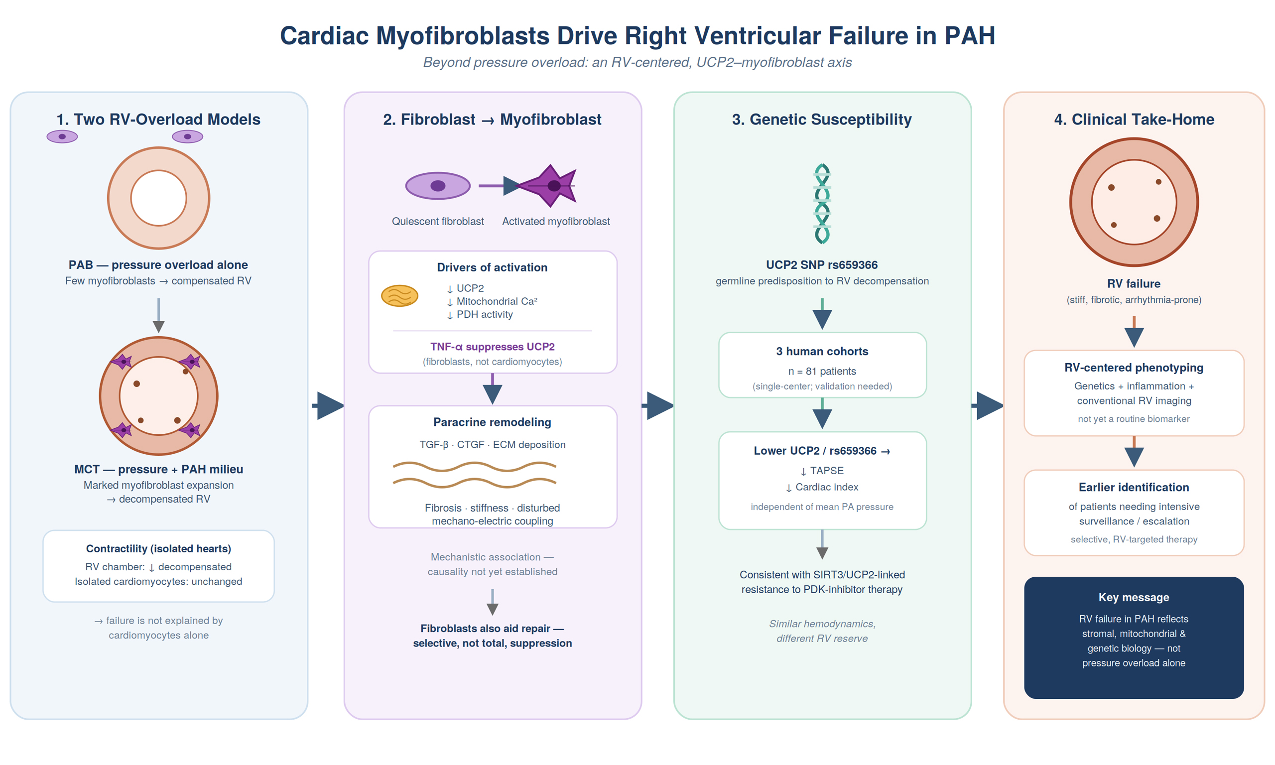
Zhang et al. combined two experimental models with three human cohorts comprising 81 patients[3]. In the monocrotaline model, decompensated RVs showed a marked increase in cardiac myofibroblasts, whereas pulmonary artery banding did not reproduce the same pattern. Unlike pulmonary artery banding, which induces isolated right ventricular pressure overload, the monocrotaline model reproduces the inflammatory and profibrotic milieu of PAH, likely explaining the greater myofibroblast expansion beyond pressure overload alone[4-7]. The authors found decreased RV contractility in isolated decompensated RV hearts, but isolated cardiomyocyte contractility remained unchanged, suggesting a non-cardiomyocyte mechanism of failure.
The study, therefore, places cardiac fibroblasts and myofibroblasts closer to the center of RV pathobiology. Cardiac fibroblasts are not merely unidimensional matrix-producing cells. Following injury or chronic stress, they can acquire a myofibroblast phenotype characterized by enhanced contractility, extracellular matrix production, and paracrine signaling[8,9]. Beyond extracellular matrix deposition, activated myofibroblasts amplify adverse remodeling through profibrotic paracrine signaling, including transforming growth factor-β and connective tissue growth factor, thereby perpetuating fibrosis and increasing myocardial stiffness. In addition, bidirectional cardiomyocyte-fibroblast crosstalk may further contribute to progressive right ventricular remodeling and dysfunction[3,6,7]. These processes may initially support structural integrity, but persistent activation and expansion can lead to stiffness, impaired relaxation, disrupted mechano-electric coupling, and increased risk of arrhythmia[9]. This interpretation should remain balanced. The data establish a strong mechanistic association across animal models, cellular experiments, and human cohorts, but do not definitively establish causality.
DISCUSSION
UCP2 and tumor necrosis factor-alpha as signals of early vulnerability
A particularly interesting aspect of the study is the proposed association between inflammation, mitochondrial calcium, pyruvate dehydrogenase (PDH) activity, and myofibroblast activation. Dromparis et al. previously implicated UCP2 in mitochondrial calcium regulation and pulmonary vascular remodeling. UCP2 deficiency can reduce mitochondrial calcium uptake, suppress mitochondrial metabolism, and create a pseudohypoxic phenotype in pulmonary artery smooth muscle cells in UCP2 knockout mice[10]. Zhang et al. observed a progressive decline in UCP2 expression and mitochondrial calcium from control to compensated and then decompensated RV fibroblasts, while tumor necrosis factor-alpha (TNF-α) reduced UCP2 expression in RV fibroblasts but not in cardiomyocytes[3]. Although the precise basis for this cell-type specificity remains unknown, it may reflect intrinsic differences in inflammatory responsiveness and metabolic adaptability between cardiac fibroblasts and cardiomyocytes. In the injured heart, cardiomyocytes primarily function as stress sensors that release danger-associated signals, whereas fibroblasts exhibit marked immunometabolic plasticity, rapidly adopting inflammatory and profibrotic phenotypes through metabolic reprogramming and myofibroblast differentiation[11,12].
These findings connect two clinically relevant aspects of PAH. First, elevated inflammatory cytokines have been associated with poor prognosis in PAH and may serve as useful prognostic biomarkers[13]. Second, the germline UCP2 single-nucleotide polymorphism rs659366 may identify patients with an intrinsic susceptibility to earlier RV decompensation. In the human cohorts, lower UCP2 levels and rs659366 were associated with worse tricuspid annular plane systolic excursion and cardiac index, even among patients with similar mean pulmonary arterial pressure[3]. The observation is consistent with a previous report showing that genetic variation affecting mitochondrial biology, especially sirtuin 3 (SIRT3) and UCP2, may influence therapeutic response in PAH by contributing to resistance to pyruvate dehydrogenase kinase inhibition therapy[14].
Clinical implications
The clinical implication is not that rs659366 or TNF-α should be incorporated into routine biomarker testing. Instead, the study proposes a framework to improve RV-centered phenotyping. Patients with similar pulmonary hemodynamics may have substantially different RV reserve because their ventricles differ at the stromal, inflammatory, and metabolic levels. Incorporating genetic predisposition, inflammatory signaling, and conventional RV measures could facilitate the identification of patients who require intensive surveillance or timely treatment escalation.
From a therapeutic perspective, current PAH therapies predominantly target the pulmonary circulation, while effective RV-specific therapies remain limited[15]. The present findings suggest targeting maladaptive fibroblast activation, restoring UCP2-related mitochondrial signaling, or interrupting the inflammatory pathway. However, fibroblasts are not simply harmful cells. They play a critical role in repair and structural integrity, meaning that complete suppression of fibroblast activity may impair adaptive repair. The therapeutic goal should be selective interruption of persistent maladaptive activation while preserving the reparative functions of cardiac fibroblasts.
Several limitations should also be acknowledged. The human cohorts included a relatively modest number of patients, predominantly from a single ethnic background, which may limit the generalizability of the genetic findings. Furthermore, although the monocrotaline model reproduces many pathological features of pulmonary arterial hypertension, species-specific differences and model-dependent biology warrant caution when translating these mechanistic observations directly to human disease. Validation in larger, ethnically diverse prospective cohorts will therefore be essential.
FUTURE SCOPE
Several questions should guide the next phase of research. The association of rs659366 and TNF-α with early decompensation requires prospective validation in larger and more diverse PAH populations. Their incremental prognostic value should be tested against established risk models and serial imaging or hemodynamic markers. Mechanistic studies should determine whether the pathway is reversible after decompensation and whether it is specific to PAH rather than a shared feature of advanced RV failure. Although Zhang et al. demonstrate that restoration of UCP2 signaling attenuates fibroblast activation and improves right ventricular remodeling in experimental PAH, whether therapeutic restoration of UCP2 can reverse established RV failure remains unknown[3,16]. Finally, larger intervention studies are warranted to determine whether targeting fibroblast metabolism or inflammatory signaling improves RV function without compromising adaptive remodeling.
CONCLUSION
Zhang et al. broaden the biological model of RV failure in PAH by integrating stromal cell identity, mitochondrial biology, inflammation, and genetic susceptibility[3]. Their findings do not yet confirm a change in routine clinical practice, but they provide a strong rationale for shifting from an afterload-centered view toward a more individualized RV-centered approach. Prospective validation of RV-specific biomarkers and therapeutic targets may enable earlier identification of patients at risk for RV failure and facilitate precision-guided interventions to preserve right ventricular function. The study opens a clinically meaningful path toward earlier recognition of vulnerable patients and, potentially, therapies directed at the failing right ventricle itself.
DECLARATIONS
Authors’ contributions
Conceived the commentary: Abdelhamid M
Performed the literature review, drafted the manuscript, critically revised the intellectual content, and approved the final version of the manuscript: Abdelhamid M, Elshabrawi MN
Availability of data and materials
Not applicable.
AI and AI-assisted tools statement
During the preparation of this manuscript, the AI tool Claude Sonnet 5 (Anthropic, released 2026-06-30) was used for language editing and to assist with the visual design and layout of the Graphical Abstract. All visual elements were reviewed, edited, and approved by the authors. The tool did not influence the study design, data collection, analysis, interpretation, or the scientific content of the work. All authors take full responsibility for the accuracy, integrity, and final content of the manuscript.
Financial support and sponsorship
None.
Conflicts of interest
All authors declared that there are no conflicts of interest.
Ethical approval and consent to participate
Not applicable.
Consent for publication
Not applicable.
Copyright
© The Author(s) 2026.
REFERENCES
1. Humbert M, Kovacs G, Hoeper MM, et al. 2022 ESC/ERS Guidelines for the diagnosis and treatment of pulmonary hypertension. Eur Heart J. 2022;43:3618-731.
2. van de Veerdonk MC, Kind T, Marcus JT, et al. Progressive right ventricular dysfunction in patients with pulmonary arterial hypertension responding to therapy. J Am Coll Cardiol. 2011;58:2511-9.
3. Zhang Y, Bonnet S, Provencher S, et al. Critical contribution of cardiac myofibroblasts in right ventricular failure and the role of UCP2 SNPs in the predisposition to RV decompensation in pulmonary arterial hypertension. Circulation. 2026;153:1494-515.
5. Aujla PK, Kassiri Z. Diverse origins and activation of fibroblasts in cardiac fibrosis. Cell Signal. 2021;78:109869.
6. McNair BD, Shorthill SK, Bruns DR. More than just a small left ventricle: the right ventricular fibroblast and ECM in health and disease. Am J Physiol Heart Circ Physiol. 2023;325:H385-97.
7. Frangogiannis NG. Fibroblasts and the extracellular matrix in right ventricular disease. Cardiovasc Res. 2017;113:1453-64.
8. Humeres C, Frangogiannis NG. Fibroblasts in the infarcted, remodeling, and failing heart. JACC Basic Transl Sci. 2019;4:449-67.
9. López B, Ravassa S, Moreno MU, et al. Diffuse myocardial fibrosis: mechanisms, diagnosis and therapeutic approaches. Nat Rev Cardiol. 2021;18:479-98.
10. Dromparis P, Paulin R, Sutendra G, Qi AC, Bonnet S, Michelakis ED. Uncoupling protein 2 deficiency mimics the effects of hypoxia and endoplasmic reticulum stress on mitochondria and triggers pseudohypoxic pulmonary vascular remodeling and pulmonary hypertension. Circ Res. 2013;113:126-36.
11. Hoque MM, Gbadegoye JO, Hassan FO, Raafat A, Lebeche D. Cardiac fibrogenesis: an immuno-metabolic perspective. Front Physiol. 2024;15:1336551.
12. Cadosch N, Gil-Cruz C, Perez-Shibayama C, Ludewig B. Cardiac fibroblastic niches in homeostasis and inflammation. Circ Res. 2024;134:1703-17.
13. Soon E, Holmes AM, Treacy CM, et al. Elevated levels of inflammatory cytokines predict survival in idiopathic and familial pulmonary arterial hypertension. Circulation. 2010;122:920-7.
14. Michelakis ED, Gurtu V, Webster L, et al. Inhibition of pyruvate dehydrogenase kinase improves pulmonary arterial hypertension in genetically susceptible patients. Sci Transl Med. 2017;9:eaao4583.
15. Prisco SZ, Thenappan T, Prins KW. Treatment targets for right ventricular dysfunction in pulmonary arterial hypertension. JACC Basic Transl Sci. 2020;5:1244-60.
Cite This Article
 Open Access
How to Cite
Download Citation
Export Citation File:
Type of Import
Tips on Downloading Citation
Citation Manager File Format
Type of Import
Direct Import: When the Direct Import option is selected (the default state), a dialogue box will give you the option to Save or Open the downloaded citation data. Choosing Open will either launch your citation manager or give you a choice of applications with which to use the metadata. The Save option saves the file locally for later use.
Indirect Import: When the Indirect Import option is selected, the metadata is displayed and may be copied and pasted as needed.
About This Article
Copyright

Data & Comments
Data
0







Comments
Comments must be written in English. Spam, offensive content, impersonation, and private information will not be permitted. If any comment is reported and identified as inappropriate content by OAE staff, the comment will be removed without notice. If you have any queries or need any help, please contact us at support@oaepublish.com.