Stimuli-responsive nanomaterials as modulators of cancer stem cell fate: current progress and future perspectives
 0
0 
Abstract
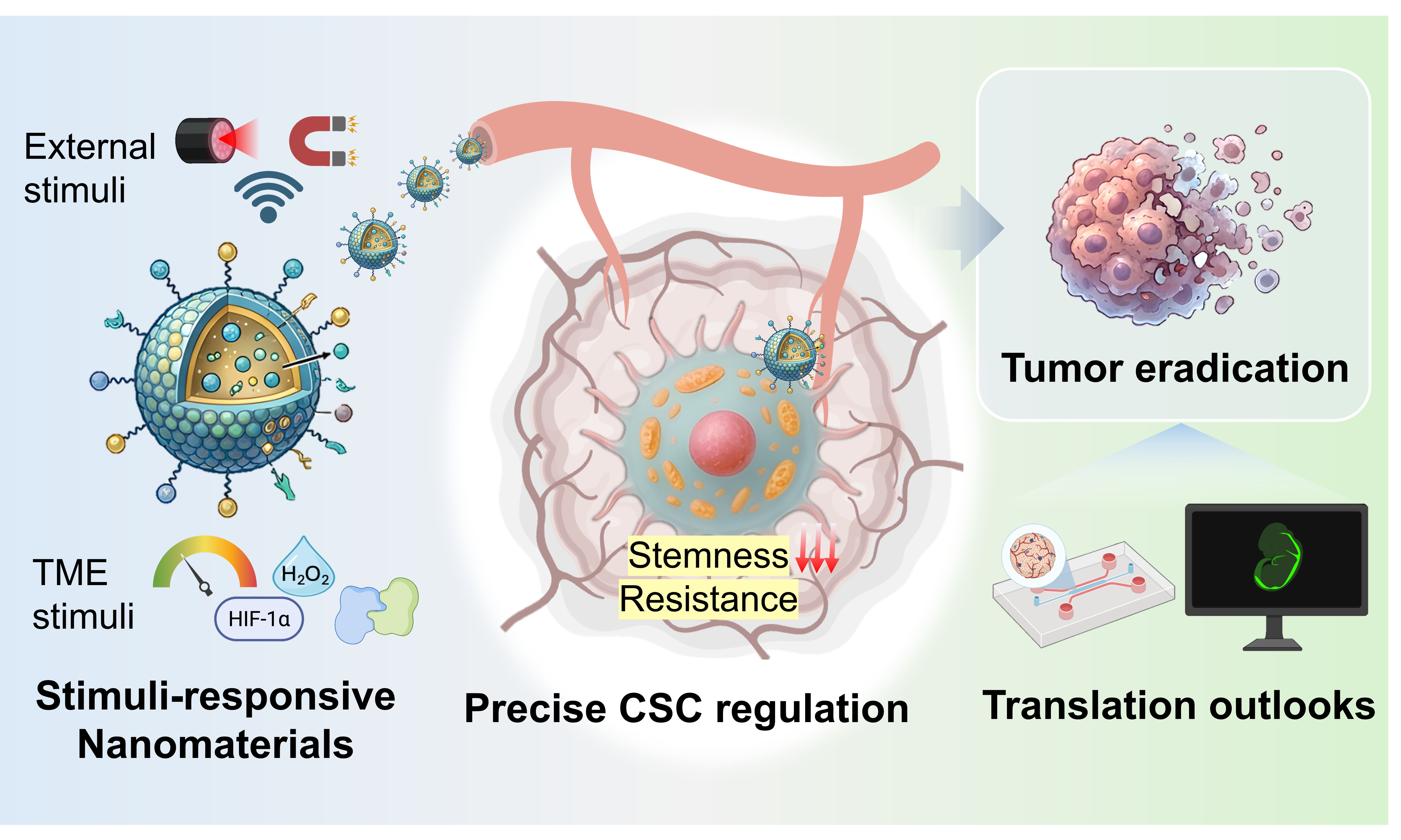
Cancer stem cells (CSCs) are the fundamental drivers of tumor metastasis, recurrence, and therapeutic resistance. They heavily rely on intricate redox homeostasis, metabolic plasticity, and specialized microenvironmental niches to maintain their stemness. This review comprehensively summarizes recent advancements in stimuli-responsive nanomaterials designed for modulating CSCs for enhanced therapeutic outcomes. Focusing on the unique biological hallmarks of CSCs, including dynamic metabolic reprogramming, a profound antioxidant defense system, and characteristic niches, we first elucidate the rational design of nanomaterials responsive to endogenous tumor microenvironment triggers and exogenous physical fields and then highlight how these smart nanoplatforms, synergized, precisely modulate cellular behaviors and stemness maintenance. Finally, we discuss pivotal translational challenges, emphasizing the urgent need for the real-time monitoring of CSCs and establishing biomimetic organoid models for the systemic interception of intercellular communications. This review provides a structural roadmap for developing next-generation multimodal nanotechnologies to achieve efficient CSC modulation and anti-tumor treatment.
Keywords
INTRODUCTION
Despite cancer stem cells (CSCs) making up only a small proportion within highly heterogeneous bulk tumors, they become the prominent driving force of tumor relapse, metastasis, and multidrug resistance due to their self-renewal and plasticity[1,2]. It has long been known that conventional chemotherapy and early targeting nanomaterials mainly focused on “killing” capacity, such as improving tumor accumulation and cytotoxicity[3]. However, these “killing” strategies severely disregard the heterogeneity and phenotype plasticity that may result in the elimination of non-CSCs but the survival of CSCs. Those residual CSCs soon differentiate and proliferate to restore malignant tumors, consequently leading to the clinical dilemma.
In the recent years, nanotechnology against CSCs is experiencing a profound conversion from simple CSC elimination to more significant CSC fate regulation. Therein, CSC elimination relies on classic cytotoxic delivery to directly induce apoptosis or necrosis in CSCs. While, fate regulation of CSCs moves beyond brute-force cytotoxicity by reprogramming cell behaviors, such as forcing phenotypic differentiation and depolarizing stemness-related networks. Compared to free drugs, stimuli-responsive nanomaterials possess superior time-space controllability, tumor retention, and on-demand dissociation characteristics, offering a multifunctional platform for precisely modulating CSCs’ behaviors[4,5]. In detail, stimuli-responsive nanomaterials advance beyond simple drug delivery systems by leveraging endogenous tumor microenvironment cues or exogenous physical fields to spatiotemporally dismantle these stemness dependencies and biomechanical barriers. Herein, this review will start by introducing the biological characteristics of CSCs, innovative design of the stimuli-responsive nanomaterials across the therapeutic paradigms, and then systematically discuss how they gradually disrupt the CSC niche and intracellular homeostasis of CSCs through physical and chemical mediation, ultimately achieving precise regulation of CSCs’ behaviors for enhanced antitumor efficacy[6].
BIOLOGICAL TRAITS OF CSCS
Biomarkers and signaling pathways. The identification and isolation of CSCs is primarily based on the overexpression of surface biomarkers, such as Prominin-1 (CD133), cluster of differentiation 44 (CD44), epithelial cell adhesion molecule (EpCAM), and CD24. Besides, intracellular enzymes or transcription factors, notably aldehyde dehydrogenase (ALDH), SRY-Box transcription factor 2 (SOX2), and octamer-binding transcription factor 4 (OCT4), are essential indicators to determine the stem-like phenotype [Table 1][7-10]. These highly expressed biomarkers not only facilitate the discrimination of CSCs, but also actively participate in maintaining stemness. For instance, CD133 is a kind of transmembrane glycoprotein that is strongly associated with tumor progression and chemoresistance[11-13]. Similarly, EpCAM (CD326) is a type of epithelial cell adhesion molecule, whose overexpression promotes signal transduction, proliferation, and differentiation of CSCs[14]. Additionally, ALDH is an intracellular enzyme that participates in oxidizing various aldehydes into their corresponding carboxylic acids, exerting a crucial role in mitigating intracellular oxidative stress levels and enhancing therapeutic resistance[15,16].
Brief summary of CSC markers, pathways, and their roles in stemness and resistance
| Category | Key marker/pathway | Role in stemness & resistance | Ref. |
| Surface markers | CD133 | Transmembrane glycoprotein; strongly associated with tumor progression and chemoresistance | [11-13] |
| CD44 | Hyaluronic acid receptor; involved in cell adhesion, migration, and resistance to apoptosis | [17] | |
| EpCAM (CD326) | Epithelial cell adhesion molecule; promotes signal transduction, proliferation, and differentiation of CSCs | [14] | |
| CD24 | Glycosylphosphatidylinositol-anchored protein; associated with metastasis and immune evasion | [18] | |
| Intracellular markers | ALDH | Detoxifies aldehydes, mitigating intracellular oxidative stress levels and enhancing therapeutic resistance | [15,16] |
| SOX2/OCT4 | Core transcription factors; maintain pluripotency and self-renewal; reprogram epigenetic landscape | [19] | |
| Signaling pathways | Wnt/β-catenin | Promotes self-renewal, differentiation, proliferation, and inhibits autophagy of CSCs; crosstalk with TGF-β promotes EMT | [20-22] |
| TGF-β | Induces EMT, immune suppression, and metastasis; cooperates with Wnt and Notch | [22] | |
| Notch | Regulates stem cell maintenance, differentiation, and chemoresistance | [23] |
CSCs frequently hyperactivate developmental signaling pathways homologous to those found in normal stem cells, and these overactivated signaling pathways are intricately interconnected, forming a complex regulatory network to promote stemness maintenance and therapeutic resistance[24]. Among these, the wingless-related integration site (Wnt) signaling cascade (including the classical Wnt signaling pathway involving β-catenin and the non-classical Wnt signaling pathway unrelated to β-catenin) has been extensively studied to interpret CSC evolution. β-catenin, as a core protein involved in signal transduction, can form a complex with T-cell factor / lymphoid enhancer-binding factor (TCF/LEF) in the nucleus to recruit cofactors to activate relevant target genes, thus promoting self-renewal, differentiation, proliferation, and inhibiting autophagy of CSCs[20,21]. Meanwhile, the Wnt signaling pathway, in conjunction with the transforming growth factor-β (TGF-β) and Notch signaling pathway, participates in the regulation of epithelial-mesenchymal transition (EMT) and serves as a key factor in promoting tumor progression[22].
Intracellular metabolism and homeostasis. Historically, CSCs were presumed to mirror the metabolic profile of differentiated bulk tumor cells, primarily relying on aerobic glycolysis (Warburg effect) to maintain energy supply. However, emerging studies reveal CSCs exhibit metabolic plasticity[25]. Unlike differentiated bulk tumor cells that rigidly rely on the Warburg effect under aerobic conditions, CSCs exhibit profound metabolic flexibility and can obtain energy by consuming oxygen and producing ATP through oxidative phosphorylation (OXPHOS), thus fully utilizing the limited nutrients in the tumor microenvironment. Beyond glucose-based energy metabolism, CSCs frequently upregulate lipogenesis, cholesterol biosynthesis, and fatty acid oxidation to meet their immense bioenergetic and membrane-building demands[26,27]. Furthermore, the accumulation of lipid intermediates actively orchestrates core stemness cascades, such as the Wnt/β-catenin and Notch signaling pathways, thereby governing self-renewal and metastatic dissemination.
In addition to metabolic characteristics, CSCs exhibit unique intracellular redox homeostasis characterized by inherently low levels of reactive oxygen species (ROS), achieved by the formation of a robust antioxidant system with elevated levels of reductive species, including glutathione and enzymes [glutathione peroxidase (GPX), superoxide dismutase (SOD), thioredoxin (TRX), etc.)[28,29]. This strengthened redox shield protects CSCs from oxidative stress, thereby conferring intrinsic resistance against ROS-based therapy (sonodynamic therapy, photodynamic therapy, ferroptosis, etc.). Concurrently, CSCs exhibit an abnormal “iron addiction” that facilitates the excessive absorption of external iron to sustain a labile “iron pool”; meanwhile, CSCs reinforce the antioxidant defense to prevent lipid peroxidation and maintain intracellular redox homeostasis[30,31]. Despite these characteristics strengthening the survival shield of CSCs to withstand external stress, this extreme reliance on metabolic and redox networks paradoxically exposes targetable vulnerabilities of CSCs for nanotherapeutic interventions[32].
The CSC niche. The spatial distribution of CSCs within solid tumors is highly heterogeneous. The majority of CSCs reside in the deep region of bulk tumors that is distant from tumor vasculature, which is acidic and extremely hypoxic. Only a minor portion of CSCs localizes within perivascular niches, exhibiting a heightened propensity for intravasation and metastatic dissemination to distant organs[33]. This highly specialized and complex microenvironment for CSCs is termed a “niche”, comprising stromal cells, immune cells, extracellular matrix (ECM), and cytokines. ECM enriched with collagen, hyaluronan and polysaccharides builds up dense physical barriers to provide mechanical support and interact with the integrin receptor of CSCs to modulate the migration and adhesion of CSCs, while cytokines secreted by other cells participate in stemness maintenance[34-37].
Paracrine signaling within this niche plays an indispensable role in supporting CSCs. Stromal cells, particularly cancer-associated fibroblast cells (CAFs), secrete transforming growth factor-β (TGF-β) to promote the self-renewal and invasive capabilities of CSCs, while vascular endothelial cells provide necessary nutrients for CSCs’ survival by secreting vascular endothelial growth factor (VEGF) and basic fibroblast growth factor (bFGF)[38]. Moreover, tumor-associated macrophages (TAMs), dendritic cells (DCs), and Regulatory T cells (Treg) would secrete immune-related cytokines and chemotactic molecules [Interleukin-10 (IL-10), C-X-C motif chemokine ligand 12 (CXCL12), TGF-β, etc.) to facilitate immune evasion of CSCs[39,40]. The CSC niche represents a formidable fortress built upon rigid ECM mechanics and corrupted stromal cellular networks; therefore, dynamically remodeling this microsystem emerges as a prerequisite for stripping CSCs of their life support.
The specialized biological traits of CSCs, as summarized in Figure 1, encompass their characteristic biomarkers and signaling pathways, intracellular metabolic and redox homeostasis, and the supportive CSC niche. Leveraging these features, researchers can rationally design smart responsive nanomaterials that can precisely modulate CSC behaviors and serve as the foundation for the following discussion.
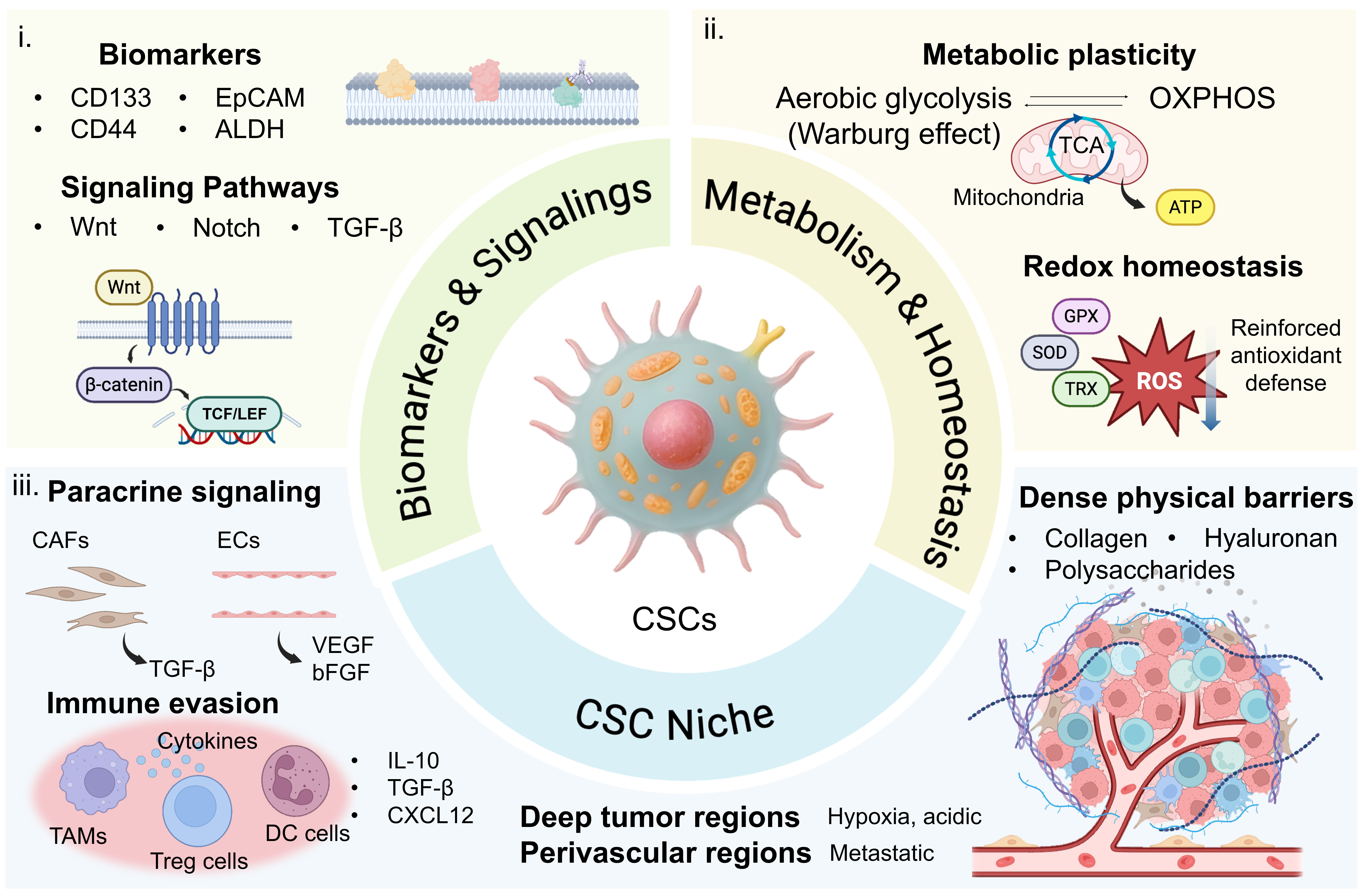
Figure 1. Schematic illustration of the specialized biological traits of CSCs. The diagram is composed of three interconnected pillars: (i) CSC-specific biomarkers and aberrant signaling pathways (e.g., CD44, CD133, ALDH, Wnt/β-catenin, and Notch), which provide molecular targets for active recognition and ligand-functionalized nanocarriers; (ii) intracellular metabolic reprogramming and redox homeostasis, characterized by enhanced glycolysis, elevated glutathione levels, and upregulated antioxidant enzymes, which offer exploitable vulnerabilities for redox responsive and metabolic-interfering nanosystems; and (iii) the CSC niche, encompassing hypoxic gradients, extracellular matrix remodeling, and stromal crosstalk, which guide the design of microenvironment-modulating nanomaterials (e.g., oxygen-replenishing, pH-sensitive, or matrix-modulating nanoplatforms). CSC: Cancer stem cell; CD133: Prominin-1; CD44: cluster of differentiation 44; EpCAM: epithelial cell adhesion molecule; ALDH: aldehyde dehydrogenase; Wnt: wingless-related integration site; TGF-β: transforming growth factor-β; TCF: T-cell factor; LEF: lymphoid enhancer-binding factor; OXPHOS: oxidative phosphorylation; TCA: tricarboxylic acid; GPX: glutathione peroxidase; SOD: superoxide dismutase; TRX: thioredoxin; ROS: reactive oxygen species; CAF: cancer-associated fibroblast cell; EC: endothelial cell; VEGF: vascular endothelial growth factor; bFGF: basic fibroblast growth factor; IL-10: Interleukin-10; CXCL12: C-X-C motif chemokine ligand 12; DC: dendritic cell; TAM: tumor-associated macrophage.
STIMULI-RESPONSIVE NANOMATERIALS: INNOVATION DESIGN
In this paradigm of modulating CSCs’ behaviors, nanomaterials no longer merely act as the passive drug delivery vehicles. Instead, their intrinsic physicochemical properties and diverse functions can serve as potent and active modulators for tumor regulation. As illustrated in Figure 2, stimuli-responsive nanomaterials used in cancer treatment are regulated by intrinsic chemical regulation and/or external stimuli-driven regulation, enabling controlled therapeutic action.
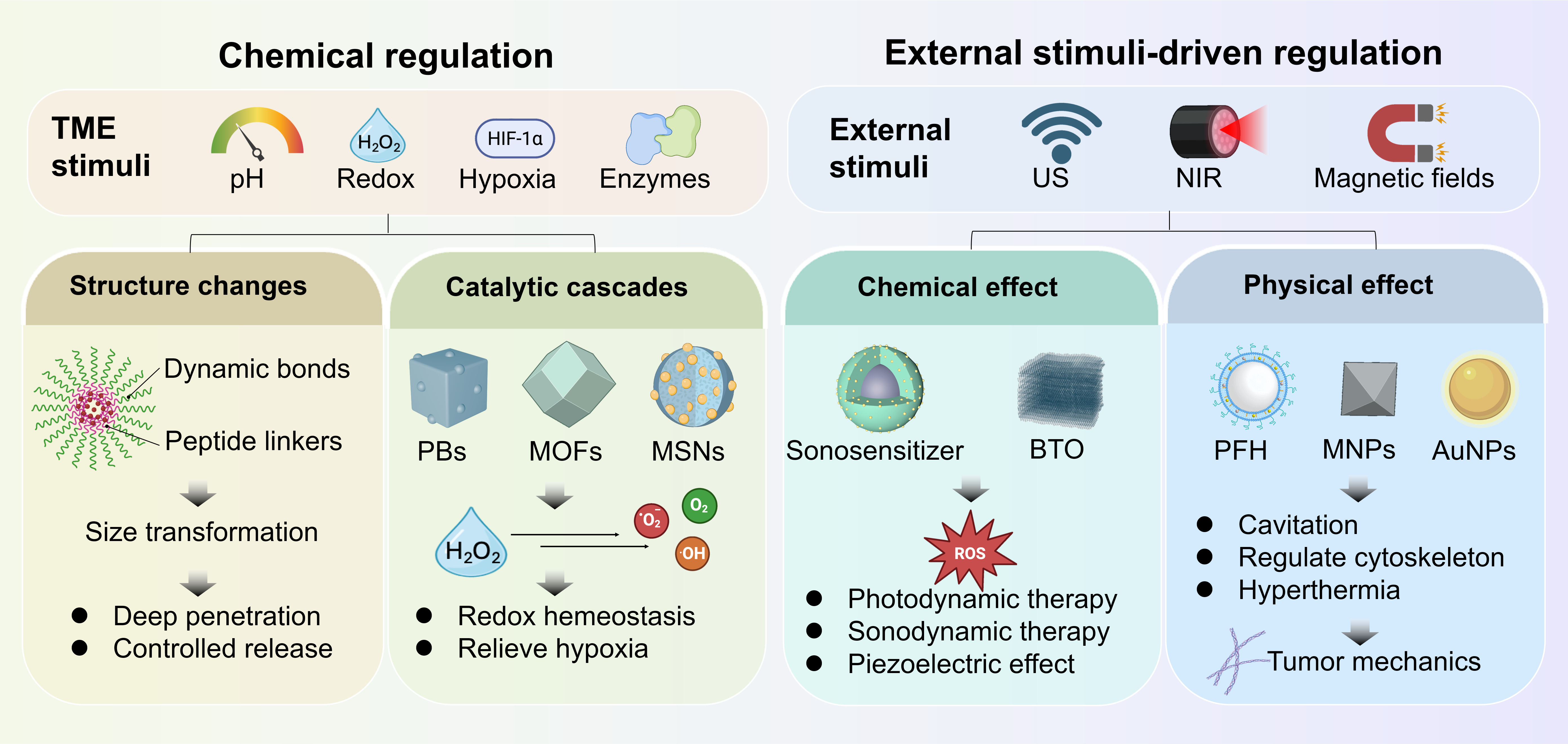
Figure 2. Schematic illustration of potential strategies by stimuli-responsive nanomaterials for modulation of CSCs. TME: Tumor microenvironment; US: ultrasound; NIR: near-infrared light; CSC: cancer stem cell; PB: Prussian blue; MOF: metal-organic framework; MSN: mesoporous silica nanoparticle; BTO: barium titanate; PFH: perfluorohexane; MNP: magnetic nanoparticles; AuNP: gold nanoparticle.
Chemical regulation. The characteristic hallmarks of the tumor microenvironment (TME) include weak acidity, elevated levels of hydrogen peroxide (H2O2), overexpressed enzymes (e.g., matrix metalloproteins, MMPs), and hypoxia, which provide endogenous triggers for stimuli-responsive chemical designs[41]. By integrating dynamic covalent bonds (e.g., disulfide bonds, boronic ester bonds) or enzyme-cleavable peptide linkers into nano-frameworks, the highly spatiotemporally controlled drug release process can be achieved[42]. For instance, pH-responsive systems utilizing protonatable polymers or acid-labile linkages ensure that chemotherapeutics or stemness inhibitors are released selectively within the acidic CSC niche, thereby enhancing local therapeutic efficacy while avoiding systemic toxicity[43]. Furthermore, pH or enzyme (MMP2) responsiveness of nanomaterials can facilitate in situ structural transformation from larger size to smaller size, thereby achieving deep tumor penetration for drug delivery and CSC elimination[44,45].
Beyond intelligent drug delivery functions, endogenous stimuli-responsive nanomaterials, particularly functional inorganic frameworks (e.g., Prussian blue analogs, metal-organic frameworks, and mesoporous silica, etc.), can interact with the TME to manifest a series of catalytic cascades[46]. By interacting with endogenous H2O2, the incorporation of metal ions (iron, cobalt, copper, etc.) and electron transfer mechanisms of nanomaterials enable the active recalibration of intracellular redox homeostasis while remodeling the hypoxia niche. For CSCs, which critically depend on adjusted redox balance to maintain stemness, the nanomaterials-induced redox turbulence can be lethal. The amplified ROS generation in CSCs disrupts core signal transductions responsive for stemness maintenance and therapeutic tolerance, thereby driving CSCs out of their protective, quiescent states and rendering them susceptible to therapeutic interventions[28].
External stimuli-driven regulation. Functional design on structure and composition endows nanomaterials with the capabilities to respond to exogenous physical fields, such as near-infrared light (NIR), ultrasound (US), alternating magnetic field, or electric field. This external stimuli-driven regulation, either through catalytic regulation (e.g., sonodynamic therapy, SDT; photodynamic therapy, PDT) or physical interruption, allows for the dynamic remodeling of TME and cellular behaviors, laying a robust material foundation for multimodal synergistic therapies. The design of NIR-responsive nanomaterials mainly involves the incorporation of noble metals with strong localized surface plasmon resonance (LSPR) effects (e.g., gold nanorods/nanostars)[47], two-dimensional transition metal dichalcogenides (e.g., MoS2 nanosheets)[48] or up-conversion nanoparticles (UCNPs)[49]. Recent advancements emphasize the deep tissue penetration of NIR-II, effectively reaching the deep-seated CSC niches. The rapid hyperthermia directly induces tumor cell apoptosis or necrosis via protein denaturation and ROS generation. Concurrently, it disrupts the ECM by denaturing dense collagen fibers and downregulating hyaluronic acid, which significantly enhances therapeutic permeability[50].
Similarly, US-mediated acoustic cavitation exerts severe mechanical stress. This mechanical stress can be amplified by the integration of fluorocarbon-encapsulated nanodroplets[51,52], which manifests significantly enhanced mechanical disruption through acoustic droplet vaporization. Rationally designed surface roughness and hydrophobicity on these nanomaterials provide optimal nucleation sites for microbubbles, elevating the cavitation yield[53,54]. At the cellular level, the violent implosion of cavitation bubbles causes transient sonoporation, increasing membrane permeability, or leads to direct mechanical lysis. Within TME, the resulting shockwaves physically shear the rigid ECM architecture, dismantling the dense stromal barrier and disrupting biomechanical supports for CSC stemness maintenance. What’s more, piezoelectric nanomaterials (e.g., barium titanate, zinc oxide) have emerged as a transformative frontier in stimuli-responsive nanomaterials owing to their unique capability of converting US-mediated mechanical stress into electric signals or ROS, severely disrupting intracellular homeostasis of tumor cells[55]. Besides, magnetic nanoparticles (MNPs), with unique magnetic properties, can dynamically convert magnetic energy into mechanical energy to regulate biological processes in response to a magnetic field[56]. MNPs conjugated with actin-binding protein (ABP-MNs) selectively induce F-actin depolymerization and reshape the tumor mechanical microenvironment, unlocking new therapeutic avenues for cancer treatment[57].
DISRUPTING ENDOGENOUS HOMEOSTASIS TO COMPROMISE CSC STEMNESS
Redox homeostasis disruption. CSCs maintain their robust self-renewal capacity, phenotypic plasticity, and therapeutic resistance by establishing a highly atypical homeostatic network characterized by a reinforced antioxidant defense system and flexible metabolic reprogramming. Targeting intracellular redox homeostasis is a primary target for such interventions, as elevated antioxidant defense maintains a significantly lower ROS level, which makes it a potential strategy for exploiting rationally designed responsive nanomaterials to dismantle CSC antioxidant shields[58]. For instance, researchers have integrated a redox-responsive dimeric prodrug of docetaxel (DTX) with the anti-CSC agent salinomycin (SAL), creating an efficient delivery system entirely devoid of inert carriers[59]. Upon endogenous cleavage, this nanosystem uniquely targets CSCs and interferes with the delicate redox homeostasis that CSCs heavily rely on for survival and chemoresistance. The above strategy capitalizes on the redox vulnerability of CSCs. Interestingly, this redox reliance is closely intertwined with another metabolic feature of CSCs, their avid demand for iron, which not only sustains their proliferative and survival capacities but also renders them susceptible to ironmediated cytotoxic reactions, most notably the Fenton chemistry.
CSCs exhibit strong addiction to iron and form a labile iron pool to meet their metabolic demands, thus making the iron-based Fenton reaction a widely utilized nanotherapeutic for CSC treatment[60]. For example, a nebulized nanocatalytic medicine (Dex@Fe-HMSNs@Ct, DFHC) was employed to modulate TAMs and effectively trigger CSC ferroptosis in the early stage of lung cancer [Figure 3A]. It has been found that DFHC-internalized TAMs localized proximal to tumor cells, thereby enabling citrate (Ct)-mediated iron transportation to trigger CSC ferroptosis by disrupting iron metabolism [Figure 3B][61]. Moreover, Wu et al. pointed out that high levels of the Prominin2 protein and GSH enabled breast CSCs to efflux excess iron ions and inhibit the generation of lipoperoxidation, thus limiting the therapeutic efficacy of ferroptosis in breast CSCs[62]. Therefore, they constructed a hyaluronic acid (HA)-modified siProminin2-loaded spindle-like FeOOH-based nanoparticle (FeOOH/siPROM2@HA) to increase the intracellular iron pool and deplete GSH, thereby effectively inducing CSC ferroptosis[62]. Similarly, a responsive nanoplatform was developed to integrate a gallic acid-modified FeOOH framework with a DKK1 inhibitor, which was found to successfully inhibit the expression of SLC7A11 and GPX4, dismantling the restoration of the antioxidant defense system and triggering a ROS storm in response to H2O2 for inducing CSC ferroptosis[63] [Figure 3C-E]. More importantly, this disruption of intracellular redox homeostasis effectively diverted CSCs away from their protected state and further downregulated the expression of stemness-associated genes, underscoring its essential role in maintaining CSC stemness.
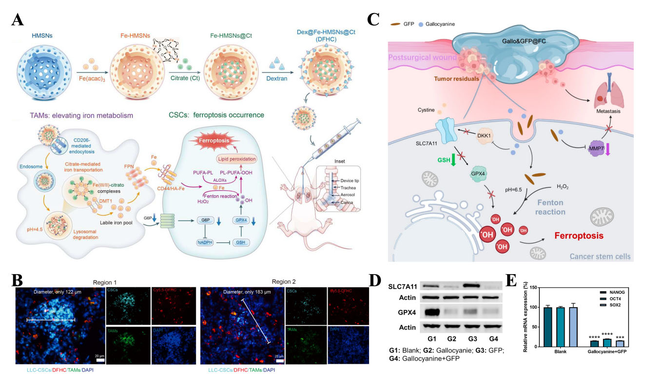
Figure 3. (A) Schematic synthetic route of DFHC and mechanisms of DFHC-regulated TAMs inducing ferroptosis of CSCs in the nebulized therapy of early orthotopic lung cancer; (B) Immunostaining results of CSCs (dark turquoise), TAMs (green), DFHC (red), and nucleus (blue, DAPI) in lung regions. Scale bar: 25 μm. Reproduced with permission[61]. Copyright 2023, American Chemical Society; (C) Schematic illustration of the ferroptosis amplification strategy to prevent tumor recurrence and metastasis after resection surgery; (D) Western blot analysis of the expression of SLC7A11 and GPX4 in CSCs under different treatments; (E) qPCR analysis of the expression of stemness-related genes in CSCs after treatment. Reproduced with permission[63]. Copyright 2024, American Chemical Society. HMSN: Hollow mesoporous silica nanoparticles; DFHC: TAM: tumor-associated macrophage; CSC: cancer stem cell; FPN: DMT: PUFA: PL: ALOX: GPX4: glutathione peroxidase 4; GSH: NADPH: GFP: FC: SLC7A11: DKK1: MMP7: NANOG: OCT4: octamer-binding transcription factor 4; SOX2: SRY-Box transcription factor 2; LLC: DAPI: qPCR:
Despite these promising effects, endogenous redox-based interventions still face inherent limitations, such as suboptimal selectivity, compensatory antioxidant upregulation, and tumor microenvironmental buffering. To address these challenges and to further augment these endogenous redox-disrupting strategies, integrating exogenous stimuli-responsive functionalities has emerged as a transformative frontier. By incorporating NIR-responsive photothermal agents or US-activated sonosensitizers into nanomedicines, researchers can exploit exogenous physical fields to radically accelerate Fenton or Fenton-like reaction kinetics by generating acoustic ROS bursts[64]. This synergistic activation not only exponentially amplifies oxidative stress within CSCs but also physically disrupts the dense stromal barriers, ensuring deep therapeutic penetration and the absolute eradication of resilient CSC niches. It is worth mentioning that such redox disruption may not be strictly CSC-specific, and off-target effects on normal stem cells or proliferating tissues remain a major concern that requires careful evaluation in future studies.
Cellular metabolism intervention. Beyond redox manipulation, targeting cellular respiration and energy metabolism is an alternative pathway to regulate CSCs. Mitochondria act as the core energetic hubs for CSCs. By designing nanomaterials capable of accumulating in mitochondria, researchers can exploit elevated mitochondrial ROS or proteotoxic stress to profoundly impair mitochondrial function, disrupting the electron transport chain and suppressing OXPHOS, ultimately precipitating a catastrophic energy crisis within the CSCs[65].
Breast CSCs were demonstrated to display enhanced dependence on glutamine (Gln), which is the critical precursor for generating NADH and FADH2 for OXPHOS[66]. Wang et al. developed a folic acid-modified nanodrug delivery system (named COHF) loaded with a Lonidamine derivative (HYL001) to inhibit glutaminolysis and reduce intracellular ROS scavengers (GSH and NADPH), which consequently blunts OXPHOS for oxygen-conserving PDT, thus disrupting the intracellular redox equilibrium within CSCs[67]. Recently, cuproptosis has been defined as a novel form of cell death, which is involved with the aggregation of lipoylated proteins and destabilization of Fe-S cluster proteins in the tricarboxylic acid (TCA) cycle, inducing proteotoxic stress and cell death[68]. Xiao et al. reported that CSCs could undergo cuproptosis upon the treatment of ROS-responsive CuET@PHF nanomedicine; therein, CuET nanocrystals were stabilized with polydopamine and hydroxyethyl starch[69]. They also emphasized the disadvantage of hypoxia in triggering mitochondrial ROS and facilitating cuproptosis, as proteins in the TCA cycle could usually be downregulated under hypoxia[69]. Therefore, enzyme mimetic nanomaterials (CAT-like) with oxygen-producing capability are potentially able to physically relieve hypoxia and amplify TCA-dependent cuproptosis.
In addition to energy metabolism, other flexible metabolic reprogramming is also a core mechanism underlying CSC-mediated therapeutic resistance. Lv et al. designed a CD276/CD133 dual-targeting biomimetic nanovesicle for the synergistic co-delivery of siMTHFD2 and manganese dioxide (MnO2) nanoparticles[70]. The smart nanovesicles targeted drug-resistant CSCs to concurrently disrupt folate-nucleotide metabolism (via siMTHFD2) and remodel aberrant γ-aminobutyric acid (GABA) metabolism via MnO2 catalysis [Figure 4A]. The disruption of GABA metabolism in CSCs reconstructed the immunosuppressive microenvironment and inhibited EMT, thus enhancing chemotherapy and immunotherapy in advanced renal cell carcinoma [Figure 4B and C].
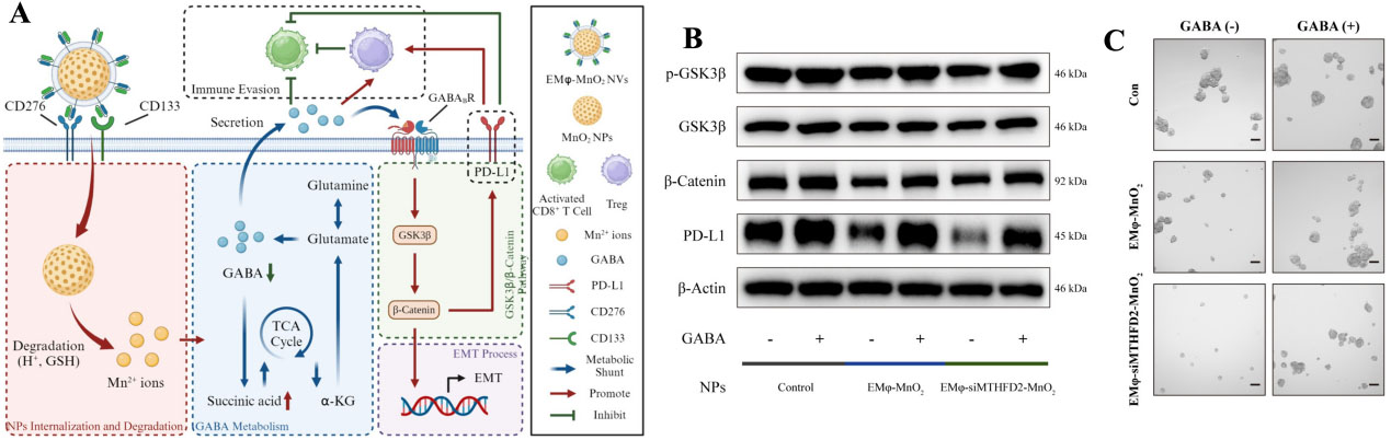
Figure 4. (A) Schematic illustration of the mechanisms of the regulatory effects on GABA metabolism induced by H-mMnO2 NVs; (B) The expression or phosphorylation rate of GSK-3β, β-Catenin, and PD-L1 in CSCs after different treatments; (C) Representative images of tumorspheres. Scale bar: 100 μm. Reproduced with permission[70]. Copyright 2024, American Chemical Society. CD: Cluster of differentiation; GABA: γ-aminobutyric acid; NV: nanovesicle; GSK-3β: Glycogen Synthase Kinase-3β; PD-L1: Programmed Death-Ligand 1; CSC: cancer stem cell; NP: nanoparticle.
REMODELING THE STROMAL MICROENVIRONMENT TO DEPRIVE CSC SUPPORT
Instead of directly targeting CSCs with cytotoxic agents, an emerging strategy involves remodeling this stromal niche to dismantle the extrinsic support system, effectively starving CSCs of the survival and self-renewal cues they require[71]. Recent advances in responsive nanomaterials offer promising strategies to remodel the tumor stromal microenvironment, thereby disrupting the external support for CSCs.
ECM remodeling. Solid tumors often exhibit severe desmoplasia, producing a dense and stiff ECM[72]. This aberrant physical environment generally serves as the biomechanical niche for protecting CSCs from external stress[73]. It forms an impenetrable barrier to nanotherapeutic delivery, and its heightened rigidity provides a mechanical scaffold that actively promotes stemness and multidrug resistance. To dismantle this mechanical shield, stimuli-responsive nanomaterials have been innovatively engineered to directly remodel the mechanics of the tumor stroma, including the intelligent release of ECM-degrading enzymes (collagenase, hyaluronidase, etc.) specifically within the tumor milieu[74,75]. This targeted enzymatic activity “softens” the stromal matrix, improving drug penetration across multiple cancer models and blocking the mechanotransductive signals (e.g., YAP/TAZ activation) that would otherwise translocate to the nucleus and drive pro-tumorigenic gene expression in CSCs. Beyond these biochemical strategies, physical regulation provides temporally and spatially controllable strategies to modify abnormal tumor mechanical properties through techniques such as mild photothermal therapy (PTT), high-intensity focused ultrasound (HIFU), hyperbaric oxygen (HBO), radiotherapy, and magneto-mechanical therapy[76]. By inducing collagen loosening and partial unfolding, these physical methods reduce the stiffness of the ECM, facilitating infiltration of nanomedicines[77]. As an example, our group has demonstrated that US-mediated mechanical effects can directly disrupt the dense ECM and reduce matrix stiffness [Figure 5A-D], thereby reducing tumor stiffness and downregulating the expression of stemness-related genes, suggesting the potential of US to remodel the biomechanical niche that supports CSC proliferation and stemness acquisition[51,64]. Physical remodeling of the ECM overcomes the desmoplastic barrier in solid tumors. These modulation approaches successfully disrupted ECM integrity, enhancing therapeutic drug penetration and reducing CSC populations. Excessive or uncontrolled ECM degradation may release matrix-sequestered growth factors (TGF-β and VEGF), which may instead promote tumor cell migration and intravasation. Besides, the proteolytic remodeling of ECM architecture can create physical corridors that facilitate CSC dissemination and metastatic outgrowth. Therefore, by integrating these strategies with spatiotemporally controlled nanomaterial systems that allow localized and on-demand ECM degradation, the risk of systemic dissemination may be mitigated.
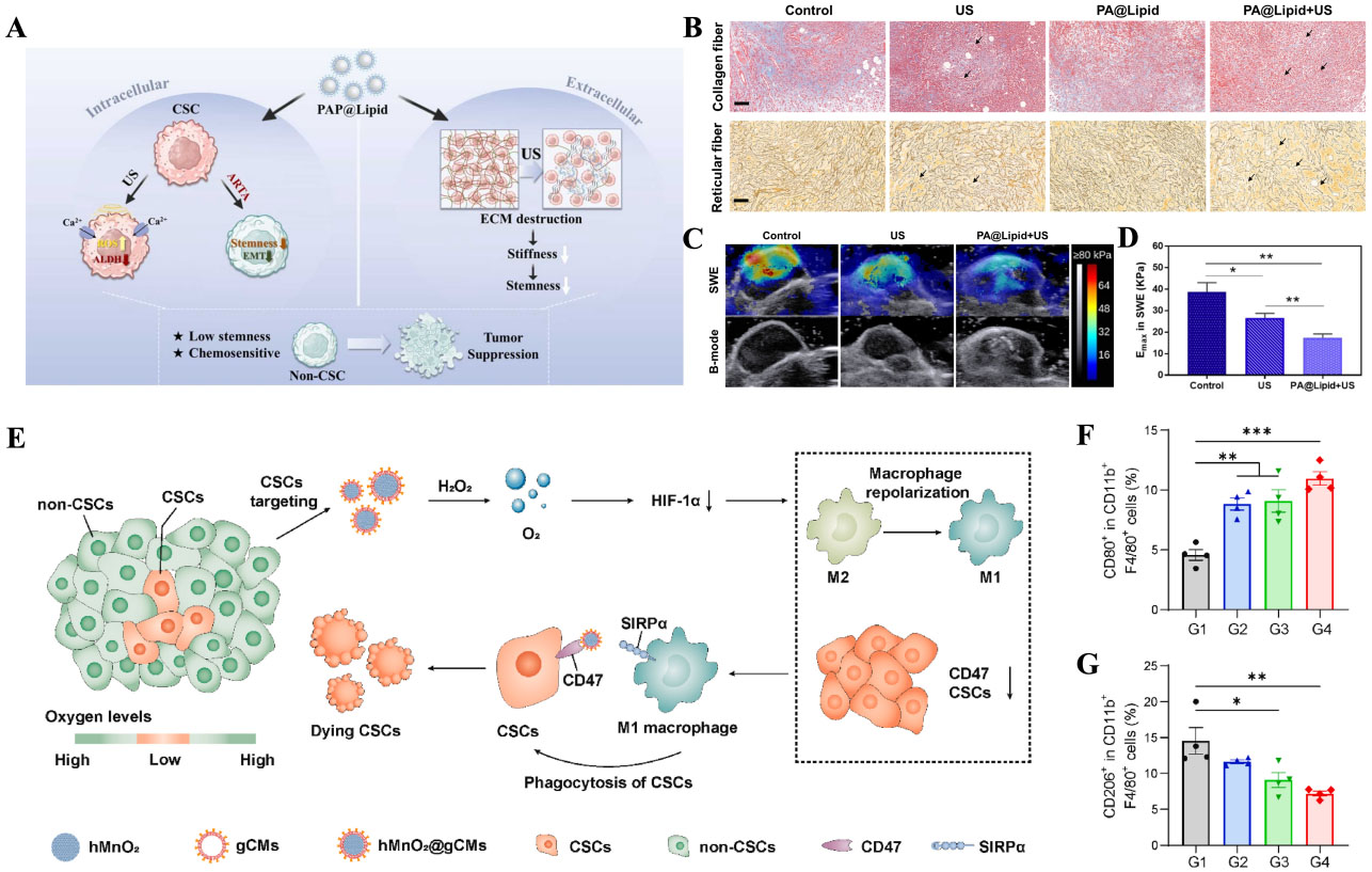
Figure 5. (A) Schematic illustrating ultrasound-mediated extracellular matrix softening to inhibit cancer stemness; (B) Tissue staining results of collagen (blue) and reticular fibers (brown) after different treatments. Scale bar: 100 μm; (C) US shear-wave elastography images of treated tumors and (D) the corresponding stiffness values presented in Young’s elastic modulus. Reproduced with permission[51]. Copyright 2024, Elsevier; (E) hMnO2@gCMs nanoparticles relieve hypoxia and promote TAMs polarization to induce robust antitumor immunity; (F) M1-like (CD80) and (G) M2-like (CD206) TAMs in tumor tissues. Reproduced with permission[95]. Copyright 2024, KeAi. CSC: Cancer stem cell; US: ultrasound; ECM: extracellular matrix; ALDH: aldehyde dehydrogenase; HIF-1α: hypoxia-inducible factor 1-alpha; CD: cluster of differentiation; TAM: tumor-associated macrophage; hMnO2@gCM: hollow manganese dioxide@cell membrane; SIRPα: signal regulatory protein α.
Reversal of hypoxia and vascular normalization. Hypoxia represents a critical sanctuary that sustains CSCs’ stemness, primarily through the constitutive activation of hypoxia-inducible factor 1-alpha (HIF-1α), which directly upregulates a panel of pluripotency-associated genes[78,79]. To alleviate this hypoxic niche, oxygen-generating nanomaterials, such as fluorocarbon-based oxygen carriers[80,81] or catalytically active manganese dioxide (MnO2)[82], have shown much promise. By remodeling the local oxygen gradient and alleviating tumor hypoxia, these responsive nanosystems effectively downregulate HIF-1α expression. This action not only directly erodes the protective external niche of CSCs but also sensitizes them to conventional therapies. For instance, Yu et al. developed a tumor hypoxia-targeting nanomedicine to improve the treatment of hypoxic cancer cells and CSCs[83]. They found that the nanomedicine could downregulate HIF-1α expression and exhibit excellent synergistic effects with paclitaxel and carboplatin, yielding ideal therapeutic outcomes[83].
As mentioned above, CSCs mainly reside in the central regions of tumor tissues that is far from tumor vasculatures, rendering them typically inaccessible due to the limited tumor-penetrating capability of nanomedicines[84]. This spatial barrier poses a particular challenge for eliminating CSCs located distantly from blood vessels. To address this issue, researchers designed a tumor-penetrating peptide (tLyP-1) conjugated ZIF-90 nanoplatform, enabling it to penetrate deeply into tumor tissues and eradicate both differentiated cancer cells and CSCs simultaneously[84]. Beyond direct penetration strategies, normalizing the tumor vasculature represents a complementary approach to enhance nanocarrier accumulation and intratumoral penetration, thereby enabling more potent eradication of CSCs. For example, Zhao et al. developed a pair of small-sized nanoparticles, sunitinib-loaded ROS-responsive micelles (RM@St) and salinomycin-loaded GSH-responsive micelles (GM@SAL)[85]. RM@St significantly extends the window of vessel normalization and enhances vessel integrity, improving the intratumoral delivery of these nanomedicines to deeply-residing CSCs, thus enhancing antitumor efficiency[85].
However, it is worth noting that vascular normalization may paradoxically increase the risk of facilitating CSC dissemination by enhancing tumor perfusion and facilitating the formation of a pre-metastatic niche. Therefore, it remains an open question whether responsive nanomaterials can effectively overcome these challenges by enabling more precisely timed and localized vascular modulation, reinforcing the promise of this strategy for CSC treatment.
Disruption of crosstalk between stromal cells and immune cells. CSCs actively co-opt surrounding stromal cells to establish a self-sustaining malignant circuit[86,87]. Through the secretion of diverse cytokines, they form reciprocal, tumor-supportive loops with CAFs and TAMs. These loops provide a constant paracrine supply of trophic factors, including interleukin-6 (IL-6) and TGF-β, which are essential for CSC maintenance[88]. Among all stromal cells, CAFs are the most abundant and play a critical role in shaping TME[89]. They not only remodel the ECM, forming a unique pathological barrier in fibrotic tumors that impedes drug delivery, but also actively interact with both immune cells and cancer cells, thereby promoting immunosuppression and CSC-mediated drug resistance[90]. In order to break this protective network, emerging nanoplatforms have been designed to target CAFs as a first-line of therapeutic intervention. Nevertheless, direct depletion of CAFs may paradoxically increase cancer aggressiveness and metastatic risk. To conquer this challenge, recent studies have developed CAFs-targeted nanoparticles that combine metabolic reprogramming with epigenetic regulation of secreted factors that promote stemness and immunosuppression. This strategy reduces CSCs and suppressor immune cell populations, thus enhancing drug sensitivity and facilitating cytotoxic T cell infiltration[86].
TAMs represent another key component of the stromal niche[91]. Among tumor-infiltrating immune cells, TAMs are generally the most abundant and have gained increasing attention due to their essential role in initiating adaptive and innate immunity, as well as their functions in pathogen defense and tumor regulation[92]. In parallel, responsive nanomaterials have been engineered to address this immune compartment, particularly through the targeted reprogramming of TAMs[93,94]. For instance, Pan et al. developed an engineered hollow manganese dioxide (hMnO2@gCMs) that generated oxygen in response to endogenous H2O2, consequently shifting TAM polarization from M2 to M1-type for potent antitumor immunity and the successful elimination of CSCs [Figure 5E-G][95]. This targeted TAM reprogramming strategy fundamentally hijacked the paracrine nutritional lines originating from the immune niche, thereby depriving CSCs of extrinsic survival signals[61,96]. By alleviating immunosuppression and disrupting CSCs’ supportive crosstalk, such combination approaches also enhance T cell infiltration and activation within the tumor bed, leading to more durable antitumor responses[97,98]. Furthermore, reducing CSC fractions via niche remodeling sensitizes CSCs to immune checkpoint blockade, addressing a major hurdle in current immunotherapy.
Collectively, stripping essential environmental support from CSCs by rationally engineered stimuli-responsive nanomaterials represents a promising strategy to overcome therapy resistance and prevent tumor relapse. ECM remodeling and vascular normalization may pose the risk of facilitating CSC dissemination. By contrast, integrating immunomodulatory functions into these nanoplatforms may offer a complementary solution, as activated immune cells can actively eliminate residual CSCs and counteract metastatic spread. Future efforts should therefore focus on combining these modalities and leveraging stromal heterogeneity to improve nanomaterial predictability and therapeutic outcomes against CSCs.
DIRECTING CSCS’ FATE VIA NANOMATERIAL INTERVENTION
Differentiation induction. Modulating intracellular homeostasis and remodeling the extracellular niche with stimuli-responsive nanomaterials are promising strategies in regulating CSCs’ behaviors (plasticity and multidrug resistance), showing significantly optimized anti-CSC and antitumor efficacy. Moreover, rationally designed stimuli-responsive nanomaterials have also emerged as sophisticated “fate programmers” by directly dictating CSC trajectories through forced differentiation, EMT reversal, and the epigenetic silencing of the core stemness network.
A commonly used paradigm in CSC regulation is nanomaterial-driven cell differentiation. By spatiotemporally delivering differentiation-inducing agents (e.g., all-trans retinoic acid, ATRA), stimuli-responsive nanomaterials can transform highly tumorigenic CSCs into a mature, differentiated, and non-stem phenotype with decreased stemness level and therapeutic tolerance[99,100]. For instance, a kind of double-layered hollow mesoporous cuprous oxide nanoparticles was developed to facilitate the sequential release of ATRA and camptothecin (CPT) within CSC niches, wherein the first release of ATRA from the outer layer induces CSC differentiation towards non-CSCs with lower chemoresistance, and then the second release of CPT from the inner layer of NPs causes cell apoptosis. Besides, the third release of Cu+ can trigger the Fenton-like reaction and glutathione depletion, resulting in ferroptosis of non-CSCs[101]. Such drug nanocarriers successfully achieve sequential drug release in response to pH changes, thereby diminishing CSC multidrug resistance and enhancing chemotherapy.
Epithelial-mesenchymal transition. Except for differentiation, reversing EMT represents a pivotal strategy to halt CSC-driven metastasis and recurrence, as EMT fundamentally drives the phenotypic plasticity, metastatic dissemination, and multidrug resistance of CSCs. It has been demonstrated that DOX increases the risk of metastasis; thus, mesoporous silica nanoparticles co-loaded with 3,3′-diindolylmethane (DIM) and DOX with exosomes as outer layers (e-DDMSNP) were synthesized[102]. The co-delivery of DIM successfully diminishes DOX-induced EMT progression at a significantly low dose and reduces CSC populations. Remarkably, fate manipulation does not strictly rely on pharmacological payloads. A hyperthermia-mediated strategy has been reported to modulate CSCs. Researchers found that targeting CD146 (EMT inducer) with black phosphorus nanosheets (BPNSs) and mild photothermal treatment was demonstrated to reverse EMT, leading to a complete stoppage of cancer cell migration [Figure 6A and B][103]. Moreover, researchers also designed CD44-modified MoS2 nanosheets to precisely target CSCs and photothermally attenuate stemness by inhibiting CD44-EMT signaling pathways, thereby reversing the EMT process and converting mesenchymal (stem-like) cancer cells to an epithelial (less stem-like) phenotype, holding great promise in addressing CSC stemness-associated challenges[104].
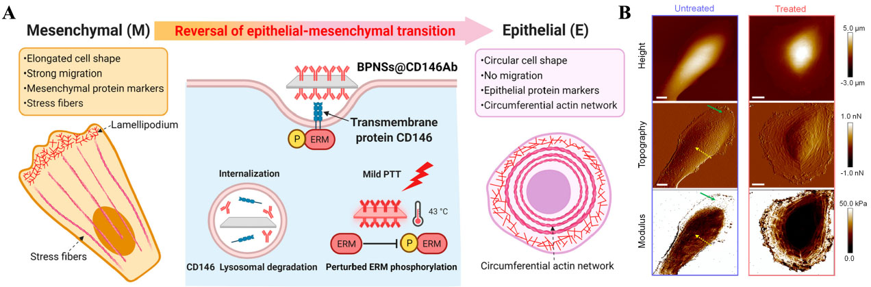
Figure 6. (A) Schematic illustration of reversing EMT in mesenchymal-type cancer cells via targeting EMT Inducer (CD146) using BPNSs and mild photothermal treatment; (B) Images of MDA-MB-231 cells in height, topography, and modulus channels after mild PTT treatment. Scale bar: 10 μm. Reproduced with permission[103]. Copyright 2022, American Chemical Society. CD: Cluster of differentiation; ERM: Ezrin, Radixin, and Moesin; PTT: photothermal therapy; EMT: epithelial-mesenchymal transition; BPNS: black phosphorus nanosheet; MDA-MB-231:M.D. Anderson-Metastatic Breast-231.
Although nanomaterials are promising in directing CSCs’ differentiating behaviors, the profound heterogeneity of CSCs across patients and tumor types severely hinders clinical translation and complicates the nanoplatform design. Rather than a one-size-fits-all approach, future designs may adopt patient-adaptable strategies guided by biomarker profiling, thereby offering a better path to balance broad utility with therapeutic efficacy.
CONCLUDING REMARKS AND OUTLOOK
In summary, the precise regulation of CSCs remains one of the most formidable challenges for cancer treatment owing to their phenotypic plasticity, intrinsic metabolic adaptability and unique protective niches. This review highlights the transformative potentials of stimuli-responsive nanomaterials in overcoming these bottlenecks. By harnessing endogenous microenvironmental cues alongside exogenous physical fields, these smart nanoplatforms may achieve spatiotemporally precise intervention. These engineered nanoplatforms can actively remodel the CSC niche and disrupt intracellular homeostasis, collectively shifting the therapeutic paradigm from merely eliminating tumor cells to systematically dismantling the foundational CSC life-support network.
Despite remarkable advances in designing stimuli-responsive nanomaterials for regulating CSCs, several critical aspects lack deeper exploration. In parallel with therapeutic efficacy, establishing precise biosafety thresholds for intense physical and biochemical stimuli, including hyperthermia, ROS production, and mechanical effects, is critical for rigorous clinical application. For instance, hyperthermia or uncontrolled ECM degradation can activate compensatory heat shock proteins or inadvertently open physical vascular escape routes, thereby accelerating tumor intravasation. Therefore, future nanomaterial designs must focus on spatiotemporally refined modulation and define therapeutic thresholds to prevent treatment-induced metastatic dissemination.
A paramount challenge in evaluating CSC-targeted therapies lies in tracking their dynamic systemic behaviors in vivo, particularly given their profound propensity for metastasis. As CSCs exhibit plasticity and undergo dynamic differentiation or EMT processes, and the extremely rare population makes monitoring their real-time differentiation trajectories and systemic dissemination a major bottleneck for current clinical non-invasive imaging modalities, including magnetic resonance imaging (MRI) or computed tomography (CT). To address this issue, functionalized stimuli-responsive nanomaterials, such as AuNPs, IONPs, or silica NPs conjugated with CSCs-specific aptamers or antibodies (e.g., CD44, CD133, ALDH), offer a promising avenue to isolate and enrich CSCs[105]. Furthermore, developing stimuli-responsive nanoprobes equipped with NIR fluorescent reporters could enable the ultra-sensitive, real-time monitoring of CSC phenotypic transitions during circulation or treatment[106]. Pioneering these in vivo tracking and dynamic capture nano systems is imperative not only for unraveling the complex temporal and spatial cascade of CSC metastasis but also for clinically intercepting systemic dissemination before secondary metastatic niches are established.
Currently, conventional preclinical models are basically standard two-dimensional (2D) cell cultures or simple 3D spheroids, which lack the complex ECM architecture, immune infiltration, and diverse cell types that are the key characteristics of the native tumor microenvironment. Thus, evaluating the distribution and therapeutic efficacy of designed nanomaterials in these oversimplified models often yields results that fail to translate to clinical treatment. To bridge this translational gap, future evaluations can integrate with advanced systems, particularly tumor organoids[107,108] or microfluidic “tumor on chip” platforms[109]. These biomimetic evaluation platforms highly preserve the functionalities and complexity of tumors, offering more reliability for assessing the regulatory effect of nanomaterials on CSC populations and their surrounding niches. Furthermore, by utilizing these platforms, excavating and intercepting the intercellular communication networks (CAFs, TAMs, etc.), and more importantly, engineering “network-disrupting” nanomaterials to regulate the “supporting cells” within niches for enhanced modalities in refractory cancer treatment[110]. Beyond biochemical signaling, biomechanical cues characterized by elevated solid stress, high interstitial fluid pressure, and ECM stiffness also serve as the fundamental driver of CSC survival and stemness maintenance. However, current research mainly focuses on disrupting ECM integrity for better penetration and more thorough CSC elimination; the underlying signaling and mechanisms between the biomechanical microenvironment and the multi-drug resistance, metastasis, and stemness traits of CSCs are worthy of further exploration.
Ultimately, while designing multifunctional, stimuli-responsive nanomaterials is theoretically promising, all preclinical strategies must confront the demands of clinical translation. At present, the vast majority of novel stimuli-responsive nanomaterials remain in the in vitro and in vivo animal testing stage. Major gaps between preclinical research and clinical trials of CSC-targeted nanotechnologies mostly lie in the long-term biosafety of nanomaterials and the dynamic heterogeneity of CSC markers in human patients. Therefore, future clinical breakthroughs will likely emerge from simpler, highly biocompatible nanomaterials with potent CSC-specific inhibitors screened and validated by patient-derived tumor organoids. By utilizing the organoid platform as a personalized screening gateway, these nanomedicines matched to individual patient biomarker profiles can finally translate responsive nanotherapeutics from the benchtop to the bedside.
DECLARATIONS
Acknowledgments
Some elements used in the Graphical Abstract and Figures 1 and 2 were created with BioRender.com
Authors’ contributions
Conception and writing: Zhu Y, Deng X
Supervision and revision: Chen H
Availability of data and materials
Not applicable.
AI and AI-assisted tools statement
During the preparation of this manuscript, the AI tool Gemini (Version 3.1 pro, released 2026-2-19) was used solely for generating some elements used in the graphical abstract and Figure 1. The tool did not influence the study design, data collection, analysis, interpretation, or the scientific content of the work. All authors take full responsibility for the accuracy, integrity, and final content of the manuscript.
Financial support and sponsorship
This work was supported by the National Natural Science Foundation of China (Grant No. 32030061), Shanghai International Cooperation Project (Grant No. 23490712900), and Sichuan Science and Technology Program (Grant No. 2026NSFSC0818).
Conflicts of interest
Chen H is an Associate Editor of Nanomedicine Therapeutics. Chen H was not involved in any steps of editorial processing, notably including reviewers’ selection, manuscript handling, and decision making. The other authors declare that there are no conflicts of interest.
Ethical approval and consent to participate
Not applicable.
Consent for publication
Not applicable.
Copyright
© The Author(s) 2026.
REFERENCES
3. Wang WD, Guo YY, Yang ZL, Su GL, Sun ZJ. Sniping cancer stem cells with nanomaterials. ACS Nano. 2023;17:23262-98.
4. Duan H, Liu Y, Gao Z, Huang W. Recent advances in drug delivery systems for targeting cancer stem cells. Acta Pharm Sin B. 2021;11:55-70.
5. Knauer N, Meschaninova M, Muhammad S, et al. Effects of dendrimer-microRNA nanoformulations against glioblastoma stem cells. Pharmaceutics. 2023;15:968.
6. Sabit H, Pawlik TM, Radwan F, et al. Precision nanomedicine: navigating the tumor microenvironment for enhanced cancer immunotherapy and targeted drug delivery. Mol Cancer. 2025;24:160.
7. Croker AK, Allan AL. Inhibition of aldehyde dehydrogenase (ALDH) activity reduces chemotherapy and radiation resistance of stem-like ALDHhiCD44+ human breast cancer cells. Breast Cancer Res Treat. 2012;133:75-87.
8. Han J, Won M, Kim JH, et al. Cancer stem cell-targeted bio-imaging and chemotherapeutic perspective. Chem Soc Rev. 2020;49:7856-78.
9. Mesrati M, Syafruddin SE, Mohtar MA, Syahir A. CD44: a multifunctional mediator of cancer progression. Biomolecules. 2021;11:1850.
10. Królewska-Daszczyńska P, Wendlocha D, Smycz-Kubańska M, Stępień S, Mielczarek-Palacz A. Cancer stem cells markers in ovarian cancer: clinical and therapeutic significance (review). Oncol Lett. 2022;24:465.
11. Behrooz A, Syahir A, Ahmad S. CD133: beyond a cancer stem cell biomarker. J Drug Target. 2019;27:257-69.
12. Cheng JX, Liu BL, Zhang X. How powerful is CD133 as a cancer stem cell marker in brain tumors? Cancer Treat Rev. 2009;35:403-8.
13. Glumac PM, LeBeau AM. The role of CD133 in cancer: a concise review. Clin Transl Med. 2018;7:18.
14. Ni J, Cozzi P, Hao J, et al. Epithelial cell adhesion molecule (EpCAM) is associated with prostate cancer metastasis and chemo/radioresistance via the PI3K/Akt/mTOR signaling pathway. Int J Biochem Cell Biol. 2013;45:2736-48.
15. Ma I, Allan AL. The role of human aldehyde dehydrogenase in normal and cancer stem cells. Stem Cell Rev Rep. 2011;7:292-306.
16. Singh S, Brocker C, Koppaka V, et al. Aldehyde dehydrogenases in cellular responses to oxidative/electrophilic stress. Free Radic Biol Med. 2013;56:89-101.
17. Chen C, Zhao S, Karnad A, Freeman JW. The biology and role of CD44 in cancer progression: therapeutic implications. J Hematol Oncol. 2018;11:64.
18. Yang Y, Zhu G, Yang L, Yang Y. Targeting CD24 as a novel immunotherapy for solid cancers. Cell Commun Signal. 2023;21:312.
19. Yang L, Shi P, Zhao G, et al. Targeting cancer stem cell pathways for cancer therapy. Signal Transduct Target Ther. 2020;5:8.
20. Krishnamurthy N, Kurzrock R. Targeting the Wnt/beta-catenin pathway in cancer: update on effectors and inhibitors. Cancer Treat Rev. 2018;62:50-60.
21. Cheng J, Tsuda M, Okolotowicz K, et al. Small-molecule probe reveals a kinase cascade that links stress signaling to TCF/LEF and Wnt responsiveness. Cell Chem Biol. 2021;28:625-635.e5.
22. Yuan X, Wu H, Han N, et al. Notch signaling and EMT in non-small cell lung cancer: biological significance and therapeutic application. J Hematol Oncol. 2014;7:87.
23. Alvarez-Trotta A, Guerrant W, Astudillo L, et al. Pharmacological disruption of the notch1 transcriptional complex inhibits tumor growth by selectively targeting cancer stem cells. Cancer Res. 2021;81:3347-57.
24. Zhou HM, Zhang JG, Zhang X, Li Q. Targeting cancer stem cells for reversing therapy resistance: mechanism, signaling, and prospective agents. Signal Transduct Target Ther. 2021;6:62.
25. Chen K, Zhang C, Ling S, Wei R, Wang J, Xu X. The metabolic flexibility of quiescent CSC: implications for chemotherapy resistance. Cell Death Dis. 2021;12:835.
26. Chen CL, Uthaya Kumar DB, Punj V, et al. NANOG Metabolically reprograms tumor-initiating stem-like cells through tumorigenic changes in oxidative phosphorylation and fatty acid metabolism. Cell Metab. 2016;23:206-19.
27. Luo X, Cheng C, Tan Z, et al. Emerging roles of lipid metabolism in cancer metastasis. Mol Cancer. 2017;16:76.
28. Diehn M, Cho RW, Lobo NA, et al. Association of reactive oxygen species levels and radioresistance in cancer stem cells. Nature. 2009;458:780-3.
29. Huang H, Zhang S, Li Y, et al. Suppression of mitochondrial ROS by prohibitin drives glioblastoma progression and therapeutic resistance. Nat Commun. 2021;12:3720.
30. Wang H, Zhang Z, Ruan S, et al. Regulation of iron metabolism and ferroptosis in cancer stem cells. Front Oncol. 2023;13:1251561.
31. Wang X, Chen Y, Wang X, et al. Stem cell factor SOX2 confers ferroptosis resistance in lung cancer via upregulation of SLC7A11. Cancer Res. 2021;81:5217-29.
32. Tuy K, Rickenbacker L, Hjelmeland AB. Reactive oxygen species produced by altered tumor metabolism impacts cancer stem cell maintenance. Redox Biol. 2021;44:101953.
33. Mohyeldin A, Garzón-Muvdi T, Quiñones-Hinojosa A. Oxygen in stem cell biology: a critical component of the stem cell niche. Cell Stem Cell. 2010;7:150-61.
34. Winkler J, Abisoye-Ogunniyan A, Metcalf KJ, Werb Z. Concepts of extracellular matrix remodelling in tumour progression and metastasis. Nat Commun. 2020;11:5120.
35. van Tienderen GS, Rosmark O, Lieshout R, et al. Extracellular matrix drives tumor organoids toward desmoplastic matrix deposition and mesenchymal transition. Acta Biomater. 2023;158:115-31.
36. Wang D, Li Y, Ge H, Ghadban T, Reeh M, Güngör C. The extracellular matrix: a key accomplice of cancer stem cell migration, metastasis formation, and drug resistance in PDAC. Cancers (Basel). 2022;14:3998.
37. Zheng X, Yu C, Xu M. Linking Tumor microenvironment to plasticity of cancer stem cells: mechanisms and application in cancer therapy. Front Oncol. 2021;11:678333.
38. Chen WJ, Ho CC, Chang YL, et al. Cancer-associated fibroblasts regulate the plasticity of lung cancer stemness via paracrine signalling. Nat Commun. 2014;5:3472.
39. Clara JA, Monge C, Yang Y, Takebe N. Targeting signalling pathways and the immune microenvironment of cancer stem cells - a clinical update. Nat Rev Clin Oncol. 2020;17:204-32.
40. Li D, Wang L, Jiang B, Jing Y, Li X. Improving cancer immunotherapy by preventing cancer stem cell and immune cell linking in the tumor microenvironment. Biomed Pharmacother. 2024;170:116043.
41. Zhang Z, Ding C, Sun T, Wang L, Chen C. Tumor therapy strategies based on microenvironment-specific responsive nanomaterials. Adv Healthc Mater. 2023;12:e2300153.
42. Wang X, Li C, Wang Y, et al. Smart drug delivery systems for precise cancer therapy. Acta Pharm Sin B. 2022;12:4098-121.
43. Zhang Z, Deng Q, Xiao C, Li Z, Yang X. Rational design of nanotherapeutics based on the five features principle for potent elimination of cancer stem cells. Acc Chem Res. 2022;55:526-36.
44. Wang Q, Cao H, Hou X, et al. Cancer stem-like cells-oriented surface self-assembly to conquer radioresistance. Adv Mater. 2023;35:e2302916.
45. Liu H, Xu S, Yong T, et al. Hydrophobicity-adaptive polymers trigger fission of tumor-cell-derived microparticles for enhanced anticancer drug delivery. Adv Mater. 2023;35:e2211980.
46. Jia X, Wang E, Wang J. Rational design of nanozymes for engineered cascade catalytic cancer therapy. Chem Rev. 2025;125:2908-52.
47. Gong J, Ding Q, Shi Y, et al. NIR-responsive gold nanomaterials in photothermal antibacterial therapy: from morphological design to multifunctional platforms. Coordin Chem Rev. 2025;526:216348.
48. Younis MR, Wang C, An R, et al. Low power single laser activated synergistic cancer phototherapy using photosensitizer functionalized dual plasmonic photothermal nanoagents. ACS Nano. 2019;13:2544-57.
49. Hu Z, Li J, Feng L, et al. Coassembly of dual-modulated AIE-ESIPT photosensitizers and UCNPs for Enhanced NIR-excited photodynamic therapy. Nano Lett. 2024;24:16426-35.
50. Yu C, Chen D, Zhu D, et al. Dual strategy of C2+ a influx and collagen denaturation to remodel the extracellular matrix and amplify sonopiezoelectric therapy. Adv Mater. 2025;37:e2501642.
51. Zhu Y, Liu T, Deng X, et al. Ultrasound-mediated intra-/extracellular dual intervening effect combined with all-trans retinoic acid for cancer stemness inhibition. Nano Today. 2024;55:102207.
52. Sheng D, Liu T, Qian L, et al. Sonodynamic and sonomechanical effect on cellular stemness and extracellular physicochemical environment to potentiate chemotherapy. J Nanobiotechnology. 2024;22:358.
53. Belova V, Gorin DA, Shchukin DG, Möhwald H. Selective ultrasonic cavitation on patterned hydrophobic surfaces. Angew Chem Int Ed Engl. 2010;49:7129-33.
54. Xu Z, Zeng Y, Kang M, et al. Dual-Enhanced sonodynamic therapy: synergizing cavitation amplification and efficient sonoluminescence-to-ROS conversion for orthotopic breast cancer treatment. Adv Mater. 2026;38:e16803.
55. Bai Y, Jiang K, Deng W, et al. Ultrasound-triggered piezoelectric biomaterials for enhanced cancer sonodynamic immunotherapy. Ultrason Sonochem. 2025;119:107402.
56. An J, Hong H, Won M, et al. Mechanical stimuli-driven cancer therapeutics. Chem Soc Rev. 2023;52:30-46.
57. Fan X, Chen H, Li Y, et al. Actin-targeted magnetic nanomotors mechanically modulate the tumor mechanical microenvironment for cancer treatment. ACS Nano. 2025;19:6454-67.
58. Mai TT, Hamaï A, Hienzsch A, et al. Salinomycin kills cancer stem cells by sequestering iron in lysosomes. Nat Chem. 2017;9:1025-33.
59. Zhao T, Chen Y, Yuan H, et al. One-stone-two-birds carrier-free nano-cocktail enables synergistic eradication of cancer cells/stem cells in breast cancer treatment. Exploration (Beijing). 2026;6:20240259.
60. Cosialls E, El Hage R, Dos Santos L, Gong C, Mehrpour M, Hamaï A. Ferroptosis: cancer stem cells rely on iron until “to die for” it. Cells. 2021;10:2981.
61. Feng Q, Fang W, Guo Y, Hu P, Shi J. Nebulized therapy of early orthotopic lung cancer by iron-based nanoparticles: macrophage-regulated ferroptosis of cancer stem cells. J Am Chem Soc. 2023;145:24153-65.
62. Wu K, Zhang W, Chen H, et al. An iron oxyhydroxide-based nanosystem sensitizes ferroptosis by a “three-pronged” strategy in breast cancer stem cells. Acta Biomater. 2023;160:281-96.
63. Zhu Y, Deng X, Dai Z, et al. A “ferroptosis-amplifier” hydrogel for eliminating refractory cancer stem cells post-lumpectomy. Nano Lett. 2024;24:8179-88.
64. Zhu Y, Deng X, Dai Z, Ning M, Miao R, Chen H. Ultrasound-orchestrated collapse of cancer stem cell niches through synergistic physical barrier disruption and intracellular homeostasis reprogramming. Biomaterials. 2026;331:124138.
65. Bonab M, Salehi S, Aghababaie A, Amini A, Alizadeh H, Behnam B. Mitochondrial hijacking and MicroRNA crosstalk: cancer stem cell-mediated immune evasion and metabolic plasticity in the tumor microenvironment. Cancers (Basel). 2026;18:1611.
66. Pacifico F, Leonardi A, Crescenzi E. Glutamine metabolism in cancer stem cells: a complex liaison in the tumor microenvironment. Int J Mol Sci. 2023;24:2337.
67. Wang Q, Li S, Xu C, et al. Glutaminolysis inhibition boosts photodynamic therapy to eliminate cancer stem cells. Biomaterials. 2024;306:122497.
68. Tsvetkov P, Coy S, Petrova B, et al. Copper induces cell death by targeting lipoylated TCA cycle proteins. Science. 2022;375:1254-61.
69. Xiao C, Wang X, Li S, et al. A cuproptosis-based nanomedicine suppresses triple negative breast cancers by regulating tumor microenvironment and eliminating cancer stem cells. Biomaterials. 2025;313:122763.
70. Lv M, Liu B, Duan Y, et al. Engineered biomimetic nanovesicles synergistically remodel folate-nucleotide and γ-aminobutyric acid metabolism to overcome sunitinib-resistant renal cell carcinoma. ACS Nano. 2024;18:27487-502.
71. Pan Y, Yuan C, Zeng C, et al. Cancer stem cells and niches: challenges in immunotherapy resistance. Mol Cancer. 2025;24:52.
72. Chitty JL, Cox TR. The extracellular matrix in cancer: from understanding to targeting. Trends Cancer. 2025;11:839-49.
73. Safaei S, Sajed R, Shariftabrizi A, et al. Tumor matrix stiffness provides fertile soil for cancer stem cells. Cancer Cell Int. 2023;23:143.
74. Xu F, Huang X, Wang Y, Zhou S. A size-changeable collagenase-modified nanoscavenger for increasing penetration and retention of nanomedicine in deep tumor tissue. Adv Mater. 2020;32:e1906745.
75. Geng M, Du H, Wei X, et al. Engineered platelets-based nano-aircraft system for precise tumor chemo-immunotherapy with graded drug delivery and self-recognized tumor targeting. Sci Bull (Beijing). 2025;70:1462-77.
76. Yang T, Wang Q, Wang L, et al. A unified biochemical-physical regulatory nanoparticle modulates tumor mechanics to augment nanomedicine penetration and antitumor efficacy. J Control Release. 2026;393:114832.
77. Chen J, Xu X, Si W, et al. Ultrasound-triggered peroxynitrite-mediated ECM degradation to enhance sonodynamic cancer therapy. Adv Healthc Mater. 2026;15:e05513.
78. Peng G, Liu Y. Hypoxia-inducible factors in cancer stem cells and inflammation. Trends Pharmacol Sci. 2015;36:374-83.
79. Lam BS, Adams GB. Blocking HIF1α activity eliminates hematological cancer stem cells. Cell Stem Cell. 2011;8:354-6.
80. Wang Y, Wang Y, Sun Y, et al. Ultrathin oleanolic acid/perfluorocarbon-ferrocene nanosheets trigger reactive species storm to reshape tumor microenvironment for enhanced chemodynamic therapy. ACS Nano. 2026;20:7831-47.
81. Ma X, Yao M, Shi J, et al. High intensity focused ultrasound-responsive and ultrastable cerasomal perfluorocarbon nanodroplets for alleviating tumor multidrug resistance and epithelial-mesenchymal transition. ACS Nano. 2020;14:15904-18.
82. Xie Y, Zhang C, Zhao Y, et al. Simultaneous modulation of hypoxia and metabolism in glioblastoma for enhanced radio-immunotherapy. Adv Funct Mater. 2024;34:2312197.
83. Yu LY, Shueng PW, Chiu HC, et al. Glucose transporter 1-mediated transcytosis of glucosamine-labeled liposomal ceramide targets hypoxia niches and cancer stem cells to enhance therapeutic efficacy. ACS Nano. 2023;17:13158-75.
84. Chen Q, Deng B, Luo Q, Song G. Deep tumor‐penetrated nanosystem eliminates cancer stem cell for highly efficient liver cancer therapy. Chem Eng J. 2021;421:127874.
85. Zhao C, Wang W, Huang Z, et al. Small-sized twin-nanoparticles normalize tumor vasculature to enhance tumor accumulation and penetration for potent eradication of cancer stem-like cells. Acta Pharm Sin B. 2025;15:5458-73.
86. Guo D, Ji X, Xie H, et al. Targeted reprogramming of vitamin B3 metabolism as a nanotherapeutic strategy towards chemoresistant cancers. Adv Mater. 2023;35:e2301257.
87. Zhong H, Zhou S, Yin S, Qiu Y, Liu B, Yu H. Tumor microenvironment as niche constructed by cancer stem cells: breaking the ecosystem to combat cancer. J Adv Res. 2025;71:279-96.
88. Liu R, Su L, Gao S, Liu W, Wang H. Cancer-associated fibroblasts functions as multifunctional architects of the tumor stroma and represent emerging therapeutic vulnerabilities. Adv Sci (Weinh). 2026;13:e10043.
89. Biffi G, Tuveson DA. Diversity and biology of cancer-associated fibroblasts. Physiol Rev. 2021;101:147-76.
90. Xue X, Wang X, Pang M, et al. An exosomal strategy for targeting cancer-associated fibroblasts mediated tumors desmoplastic microenvironments. J Nanobiotechnology. 2024;22:196.
91. Bai R, Li Y, Jian L, Yang Y, Zhao L, Wei M. The hypoxia-driven crosstalk between tumor and tumor-associated macrophages: mechanisms and clinical treatment strategies. Mol Cancer. 2022;21:177.
92. Yuan H, Qiu Y, Mei Z, et al. Cancer stem cells and tumor-associated macrophages: Interactions and therapeutic opportunities. Cancer Lett. 2025;624:217737.
93. Yang Z, Luo Y, Yu H, et al. Reshaping the tumor immune microenvironment based on a light-activated nanoplatform for efficient cancer therapy. Adv Mater. 2022;34:e2108908.
94. Jiang M, Li X, Zhang J, et al. Dual Inhibition of endoplasmic reticulum stress and oxidation stress manipulates the polarization of macrophages under hypoxia to sensitize immunotherapy. ACS Nano. 2021;15:14522-34.
95. Pan Y, Yu L, Liu L, et al. Genetically engineered nanomodulators elicit potent immunity against cancer stem cells by checkpoint blockade and hypoxia relief. Bioact Mater. 2024;38:31-44.
96. Zhang T, Wei Y, Liu Z, et al. In situ engineered “cascade-amplified” drug-loaded vesicles for enhanced cancer stem cell therapy. J Extracell Vesicles. 2026;15:e70292.
97. Gao S, Liu M, Zhang Y, et al. A precision intelligent nanomissile for inhibiting tumor metastasis, boosting energy deprivation and immunotherapy. Biomaterials. 2025;315:122953.
98. Wu Y, Diao P, Peng Y, et al. A hybrid manganese nanoparticle simultaneously eliminates cancer stem cells and activates STING pathway to potentiate cancer immunotherapy. ACS Nano. 2025;19:12237-52.
99. Shen S, Xu X, Lin S, et al. A nanotherapeutic strategy to overcome chemotherapeutic resistance of cancer stem-like cells. Nat Nanotechnol. 2021;16:104-13.
100. Pan Y, Ma X, Liu C, et al. Retinoic acid-loaded dendritic polyglycerol-conjugated gold nanostars for targeted photothermal therapy in breast cancer stem cells. ACS Nano. 2021;15:15069-84.
101. Li Z, Yang J, Ren B, et al. Double-layered hollow mesoporous cuprous oxide nanoparticles for double drug sequential therapy of tumors. Adv Mater. 2024;36:e2313212.
102. Sarkar R, Biswas S, Ghosh R, et al. Exosome-sheathed porous silica nanoparticle-mediated co-delivery of 3,3'-diindolylmethane and doxorubicin attenuates cancer stem cell-driven EMT in triple negative breast cancer. J Nanobiotechnology. 2024;22:285.
103. Liu J, Smith S, Wang C. Reversing the epithelial-mesenchymal transition in metastatic cancer cells using CD146-targeted black phosphorus nanosheets and a mild photothermal treatment. ACS Nano. 2022;16:3208-20.
104. Liu J, Smith S, Wang C. Photothermal attenuation of cancer cell stemness, chemoresistance, and migration using CD44-targeted MoS2 nanosheets. Nano Lett. 2023;23:1989-99.
105. Yun Y, Kim S, Lee SN, Cho HY, Choi JW. Nanomaterial-based detection of circulating tumor cells and circulating cancer stem cells for cancer immunotherapy. Nano Converg. 2024;11:56.
106. Li B, Tian J, Zhang F, et al. Self-assembled aldehyde dehydrogenase-activatable nano-prodrug for cancer stem cell-enriched tumor detection and treatment. Nat Commun. 2024;15:9417.
107. Zheng F, Lan Z, Chen H, Ma M. Engineering bone-mimetic microspheres to recapitulate the tumor microenvironment for in vitro osteosarcoma modeling. Biomedicines. 2026;14:868.
108. Ravi K, Manoharan TJM, Wang KC, Pockaj B, Nikkhah M. Engineered 3D ex vivo models to recapitulate the complex stromal and immune interactions within the tumor microenvironment. Biomaterials. 2024;305:122428.
109. Hu W, Bei HP, Jiang H, et al. DLM-GelMA/tumor slice sandwich structured tumor on a chip for drug efficacy testing. Lab Chip. 2024;24:3718-27.
Cite This Article
 Open Access
How to Cite
Download Citation
Export Citation File:
Type of Import
Tips on Downloading Citation
Citation Manager File Format
Type of Import
Direct Import: When the Direct Import option is selected (the default state), a dialogue box will give you the option to Save or Open the downloaded citation data. Choosing Open will either launch your citation manager or give you a choice of applications with which to use the metadata. The Save option saves the file locally for later use.
Indirect Import: When the Indirect Import option is selected, the metadata is displayed and may be copied and pasted as needed.
About This Article
Copyright

Data & Comments
Data
0




Comments
Comments must be written in English. Spam, offensive content, impersonation, and private information will not be permitted. If any comment is reported and identified as inappropriate content by OAE staff, the comment will be removed without notice. If you have any queries or need any help, please contact us at support@oaepublish.com.