


fig10
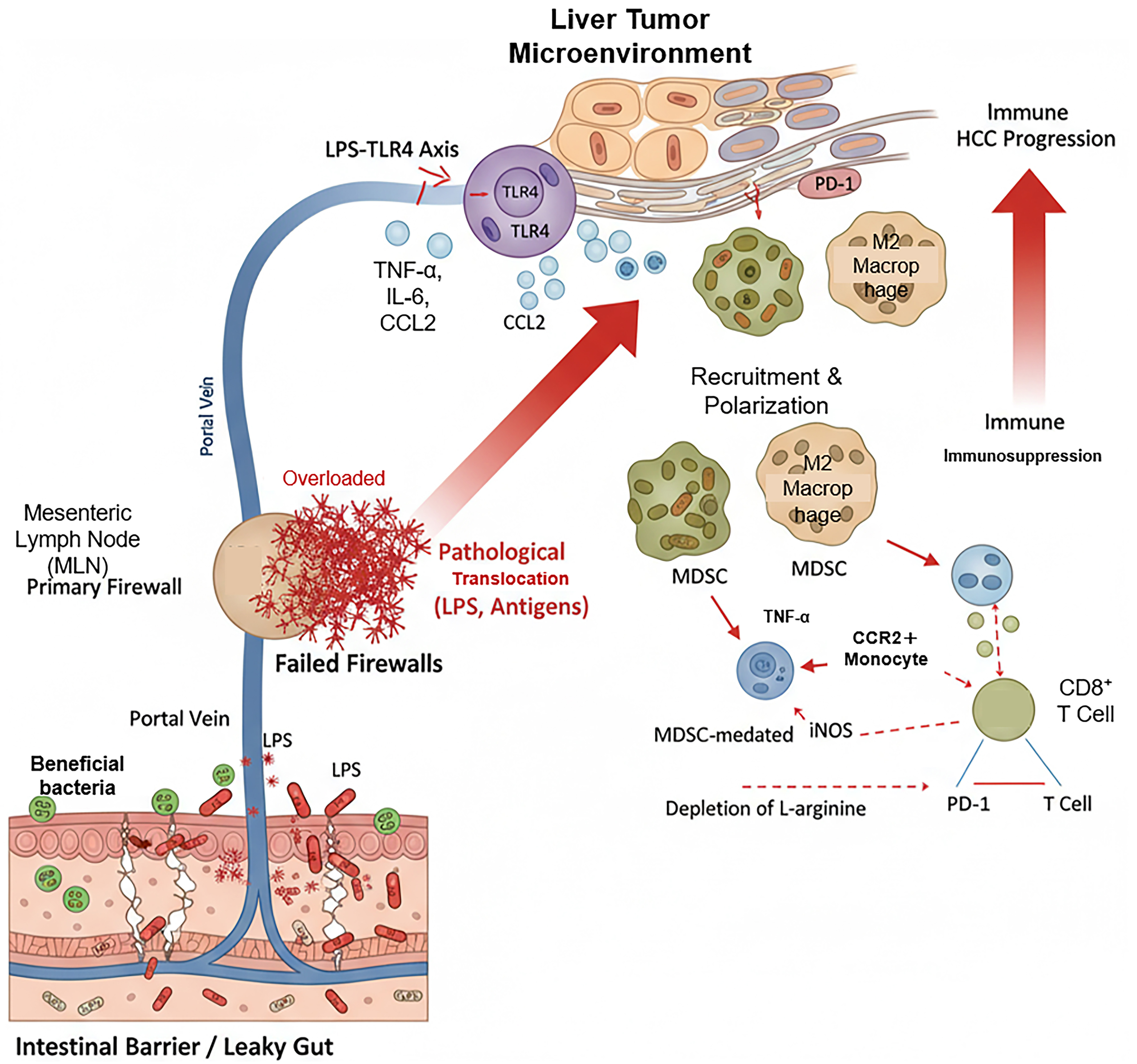
Figure 10. Mechanisms of intestinal barrier dysfunction and the “failed firewall” driving hepatic immunosuppression in HCC. The schematic illustrates the pathological translocation of LPS via the portal vein and the subsequent hyperactivation of the hepatic LPS-TLR4 axis. Solid red arrows indicate pro-inflammatory signaling, cellular recruitment, and polarization toward immunosuppressive phenotypes (e.g., MDSCs, M2 macrophages); dashed red arrows represent mechanisms of effector cell suppression (e.g., L-arginine depletion, iNOS); red T-bars denote direct functional inhibition of CD8+ T cells (e.g., via the PD-1/PD-L1 axis). Detailed descriptions of the primary and secondary immunological firewalls are provided in the corresponding text. HCC: Hepatocellular carcinoma; LPS: lipopolysaccharide; MDSCs: myeloid-derived suppressor cells; iNOS: inducible nitric oxide synthase; MLN: mesenteric lymph node; LPS-TLR4: lipopolysaccharide-Toll-like receptor 4; PD-1: programmed cell death protein 1; PD-L1: programmed death-ligand 1; TNF-α: tumor necrosis factor alpha; IL-6: interleukin 6; CCL2: C-C motif chemokine ligand 2; CCR2+: C-C motif chemokine receptor 2 positive; CD8+: cluster of differentiation 8 positive.


