


fig3
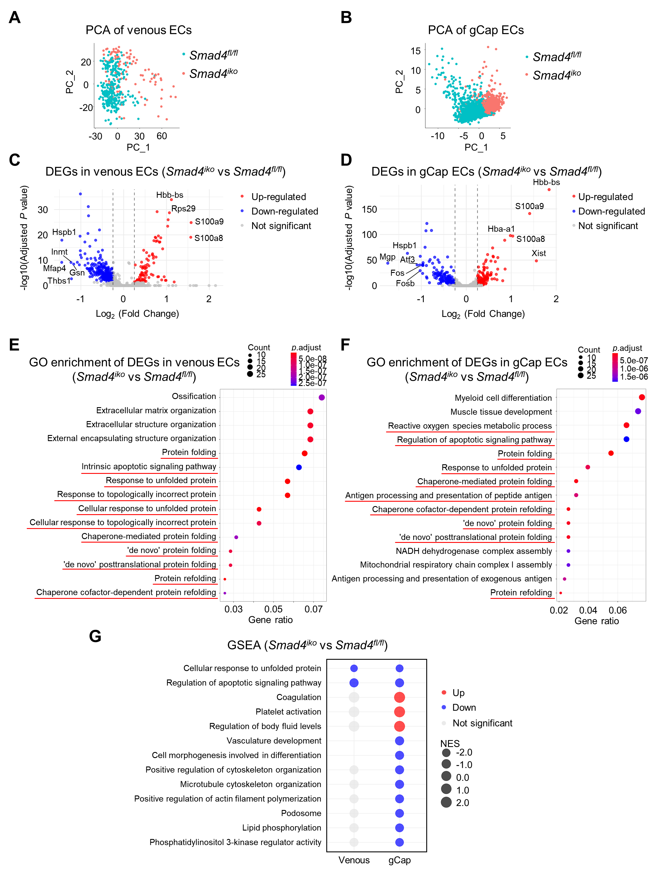
Figure 3. Characterization of the impact of Smad4 deletion on single-cell transcriptomics of pulmonary venous and gCap ECs. (A and B) Principal component analysis (PCA) of pulmonary venous ECs (A) and gCap ECs (B) from Smad4fl/fl and Smad4iko mice. Note that ECs from the two genotypes show a clear separation in both venous and gCap compartments, indicating genotype-associated transcriptomic differences. (C and D) Volcano plots of DEGs in Smad4iko vs. Smad4fl/fl pulmonary venous and gCap ECs. Significantly upregulated and downregulated genes are labeled in red and blue. Gene symbols indicate the top DEGs. (E and F) GO enrichment analysis of DEGs from pulmonary venous and gCap ECs. Note that a number of protein folding-related processes and apoptotic pathways are enriched in both EC subpopulations (red lines), indicating a common effect of SMAD4 on the maintenance of proteostasis and survival in these cells. (G) GSEA showing the indicated upregulated and downregulated pathways in Smad4iko vs. Smad4fl/fl pulmonary venous and gCap ECs. NES: normalized enrichment score. Note that Smad4iko gCap ECs show additionally upregulated pathways involved in coagulation, platelet activation and fluid homeostasis but downregulated pathways associated with vascular morphogenesis, cell morphology and cytoskeletal regulation, and phospholipid/PI3K signaling.


