


fig2
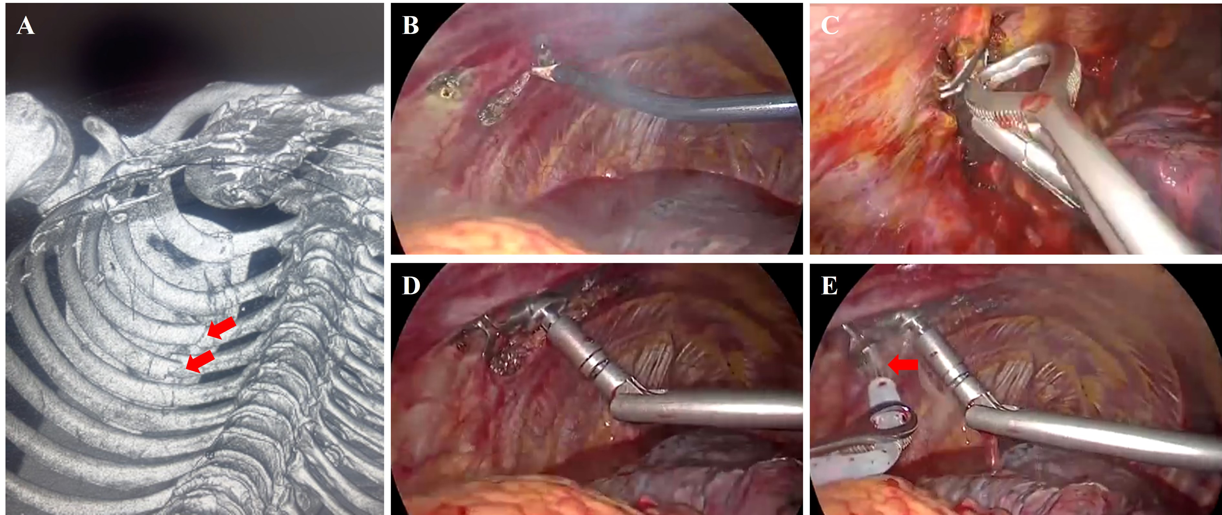
Figure 2. Intraoperative images of Complete Thoracoscopy group. (A) Preoperative three-dimensional reconstruction and fracture localization, the site of rib fracture and the fracture fragments were indicated by the red arrow; (B) Exposure of the fracture ends using an electrocautery hook; (C) “Heart-shaped” reduction forceps; (D) Specialized thoracoscopic rib plate applicator; (E) Application of sterile warm saline (indicated by the red arrow) to activate the shape memory alloy embracing fixator.


