


fig3
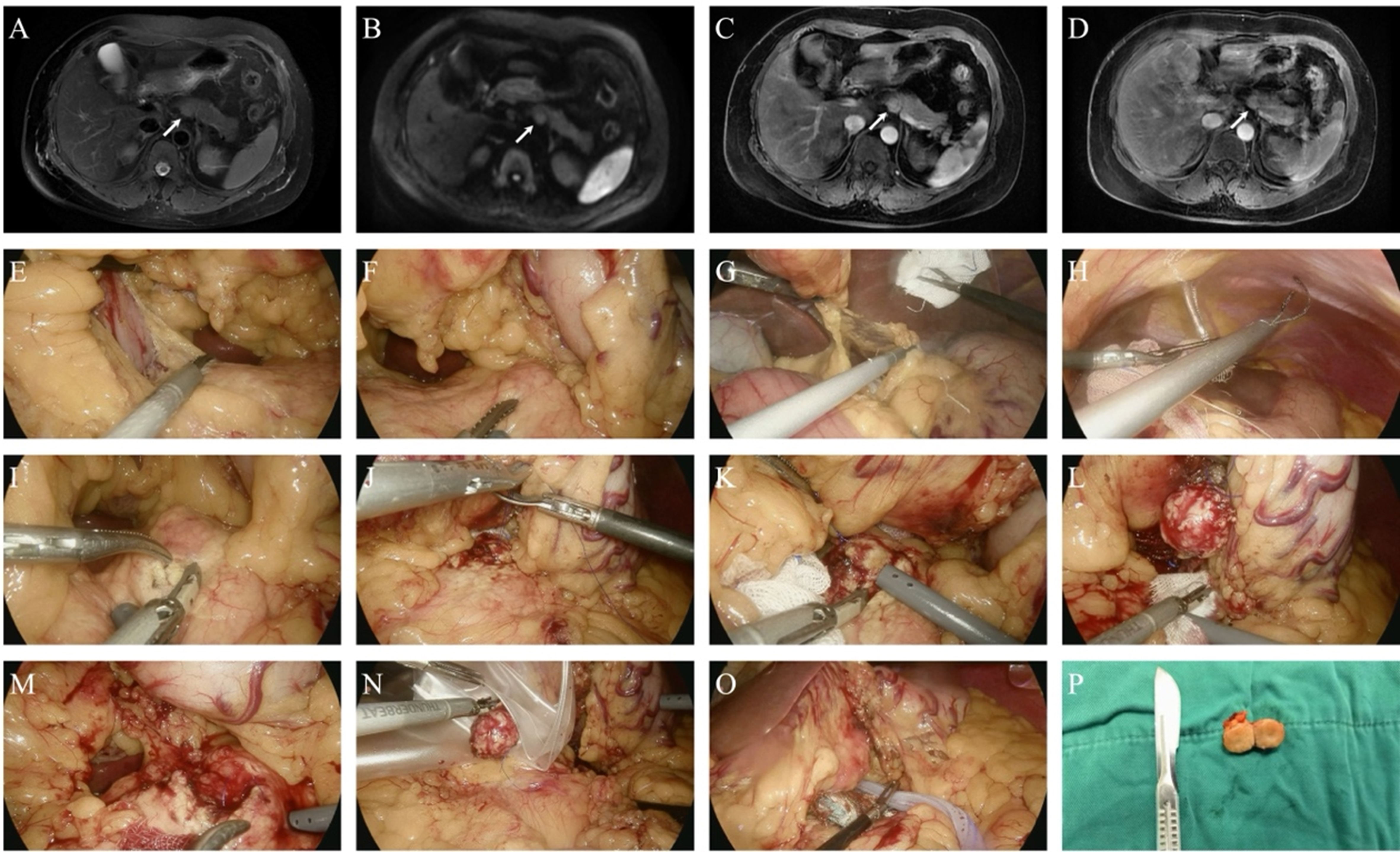
Figure 3. Representative minimally invasive enucleation of a pancreatic neuroendocrine tumor. (A-D) Preoperative magnetic resonance imaging demonstrating a well-circumscribed lesion located at the superior border of the pancreatic body, measuring approximately 1.7 cm in diameter, with slightly hyperintense signal on T2-weighted imaging, mild diffusion restriction on DWI, hyperenhancement in the arterial phase, isoenhancement in the venous phase, and no pancreatic duct dilation; (E) Adhesiolysis of the posterior gastric wall; (F) Identification of the tumor location; (G) Opening of the lesser sac; (H) Suspension of the stomach to the upper abdominal wall; (I) Incision of the pancreatic capsule along the tumor margin; (J) Suturing and suspension of the tumor; (K) Continued dissection of the lesion; (L) Complete enucleation of the tumor; (M) Ligation of arterial bleeding; (N) Specimen retrieval; (O) Placement of hemostatic materials and drainage; (P) Gross appearance of the resected specimen. DWI: Diffusion-weighted imaging.