

fig3
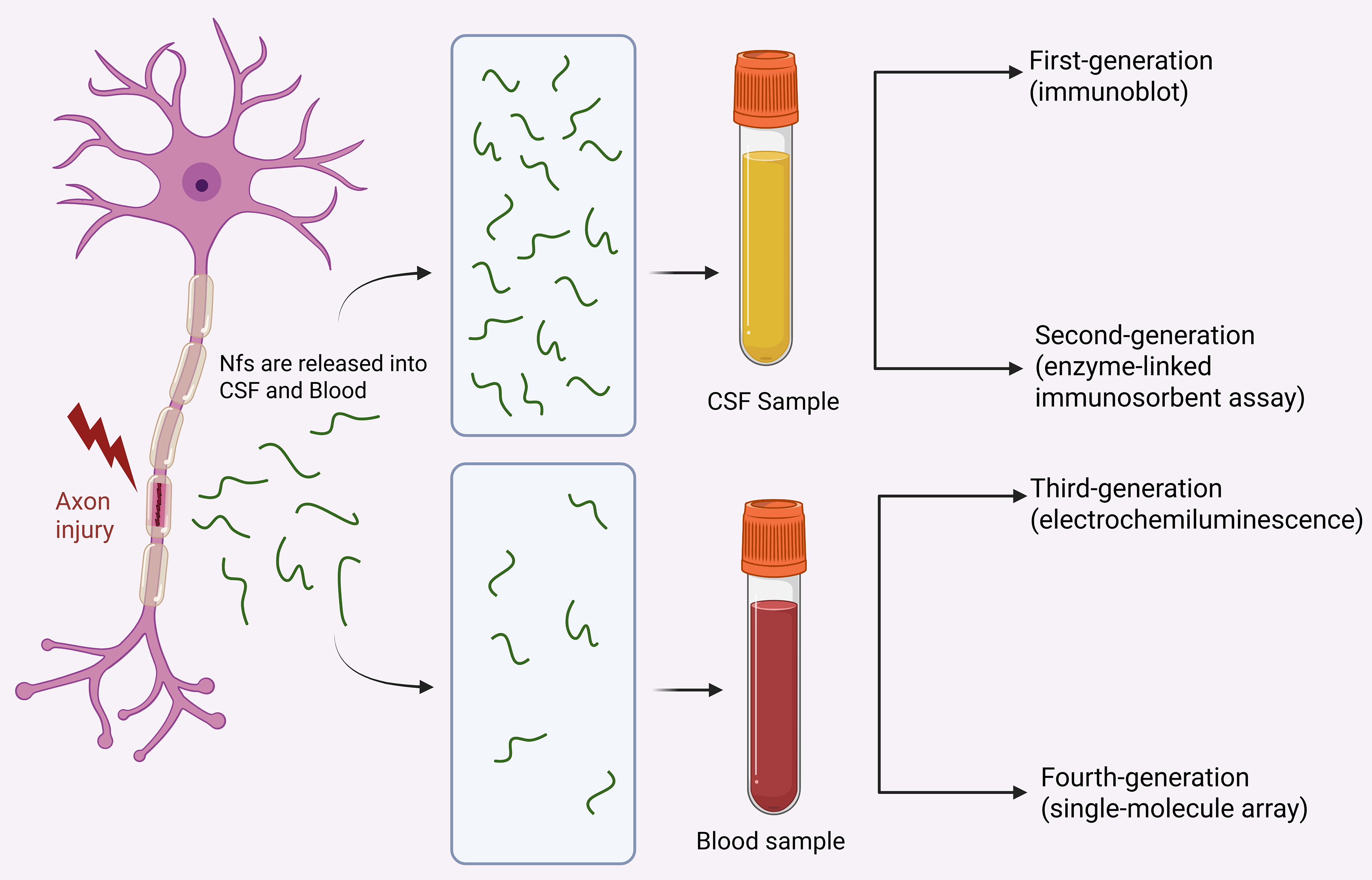
Figure 3. Release and detection of NFs after axonal injury. This figure was designed by the authors using BioRender. The schematic diagram shows that NF proteins are released into CSF and peripheral blood upon axonal damage in neurodegenerative conditions, and summarizes the evolutionary trajectory of detection technologies from early immunoblotting to ultrasensitive SiMoA assays. Created in BioRender. Zhao, X. (2026) https://BioRender.com/e86ov2c. NFs: Neurofilaments; CSF: cerebrospinal fluid; SiMoA: single-molecule array.