


fig4
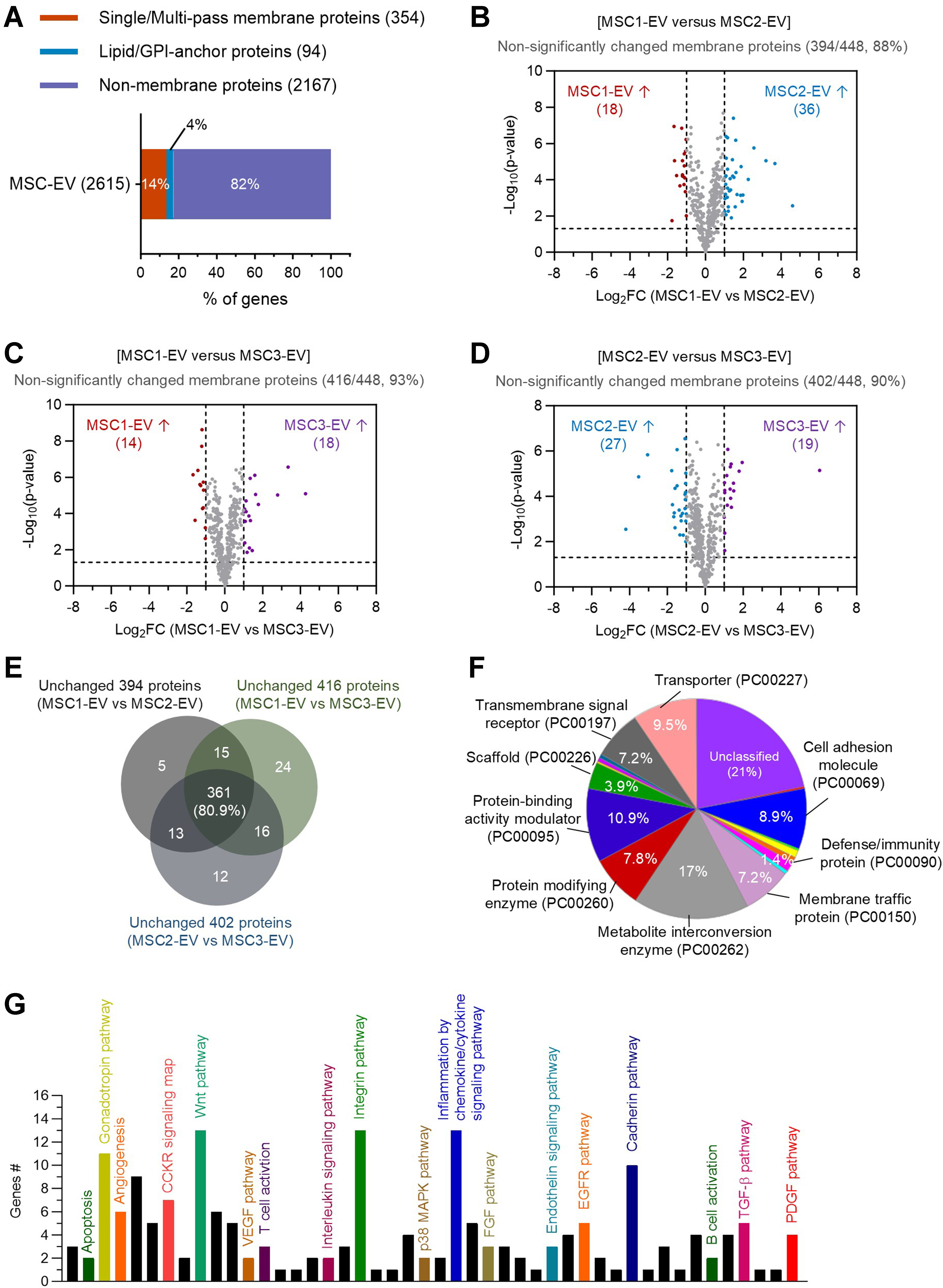
Figure 4. Analysis of conserved EV membrane proteins across donor batches. (A) Composition of membrane proteins in three MSC donor-derived EV proteome, including transmembrane proteins (354, 14%) and lipid/GPI-anchored proteins (94, 4%) of the 2,615 total proteins; (B-D) Volcano plots comparing membrane proteins between different EV batches: MSC1-EV vs. MSC2-EV (B; 394 proteins unchanged, 88%), MSC1-EV vs. MSC3-EV (C; 416 proteins unchanged, 93%), and MSC2-EV vs. MSC3-EV (D; 402 proteins unchanged, 90%); (E) Venn diagram analysis showing overlap of unchanged membrane proteins across the three pairwise comparisons, highlighting 361 proteins consistently stable across different batches; (F) Protein class analysis of the 361 unchanged membrane proteins, annotated by pathway-based functional categories; (G) Panther pathway analysis of the 361 unchanged membrane proteins, identifying signaling pathways potentially associated with EV function. EV: Extracellular vesicle; MSC: mesenchymal stem cell; GPI: glycosylphosphatidylinositol.


