





fig3
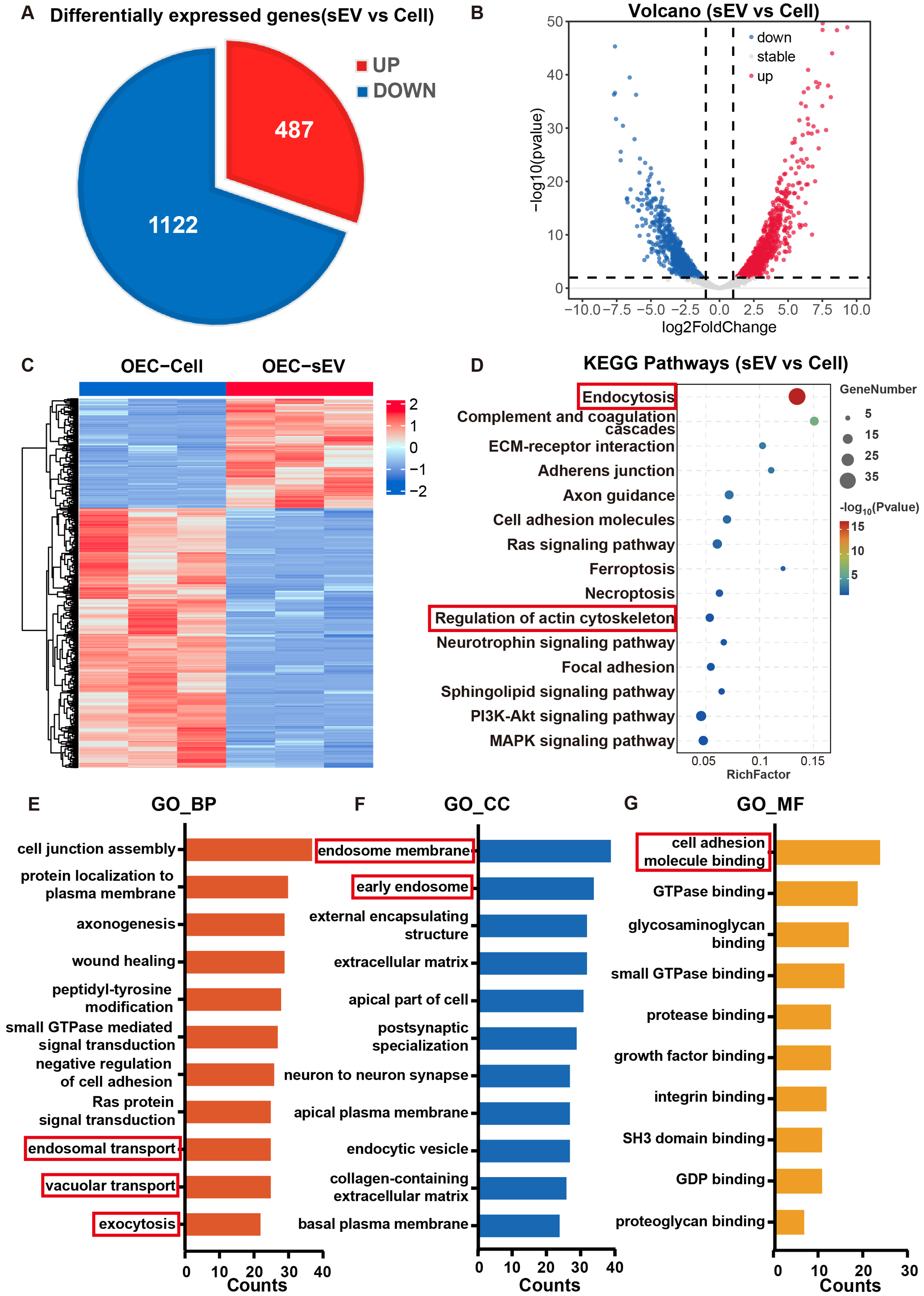
Figure 3. Proteomic profiling of OEC-sEVs vs. OEC whole-cell lysates. (A) Pie chart of differentially expressed proteins; (B) Volcano plot highlighting upregulated (red) and downregulated (blue) proteins in OEC-sEVs; (C) Heatmap of protein expression profiles; (D) KEGG analysis of highly expressed proteins in the OEC-sEV group; (E) GO (Biological Process) analysis of highly expressed proteins in the OEC-sEV group; (F) GO (Cellular Component) analysis of highly expressed proteins in the OEC-sEV group; (G) GO (Molecular Function) analysis of highly expressed proteins in the OEC-sEV group. n = 3 biologically independent OEC cultures and n = 3 biologically independent sEV preparations. Red boxes highlight the critical pathways associated with RPE phagocytosis. GO: Gene Ontology; KEGG: Kyoto Encyclopedia of Genes and Genomes; OEC: olfactory ensheathing cell; OEC-sEVs: olfactory ensheathing cell-derived small extracellular vesicles; RPE: retinal pigment epithelium; sEV: small extracellular vesicle; sEVs: small extracellular vesicles.




