

fig5
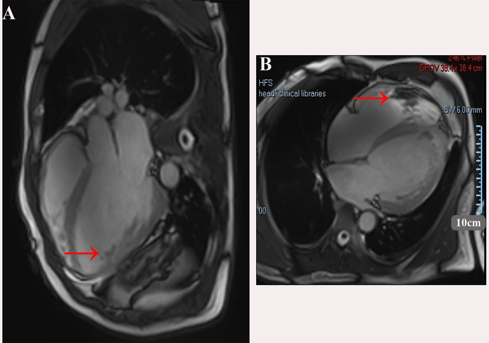
Figure 5. Showing the Cardiac magnetic resonance imaging, balanced steady-state free precession (bSSFP) cine sequence. (A) four-chamber view; (B) short-axis view. A large thrombus is seen adjacent to the apical/anterior wall of the left ventricle (red arrows). The left ventricle is markedly dilated.