


fig10
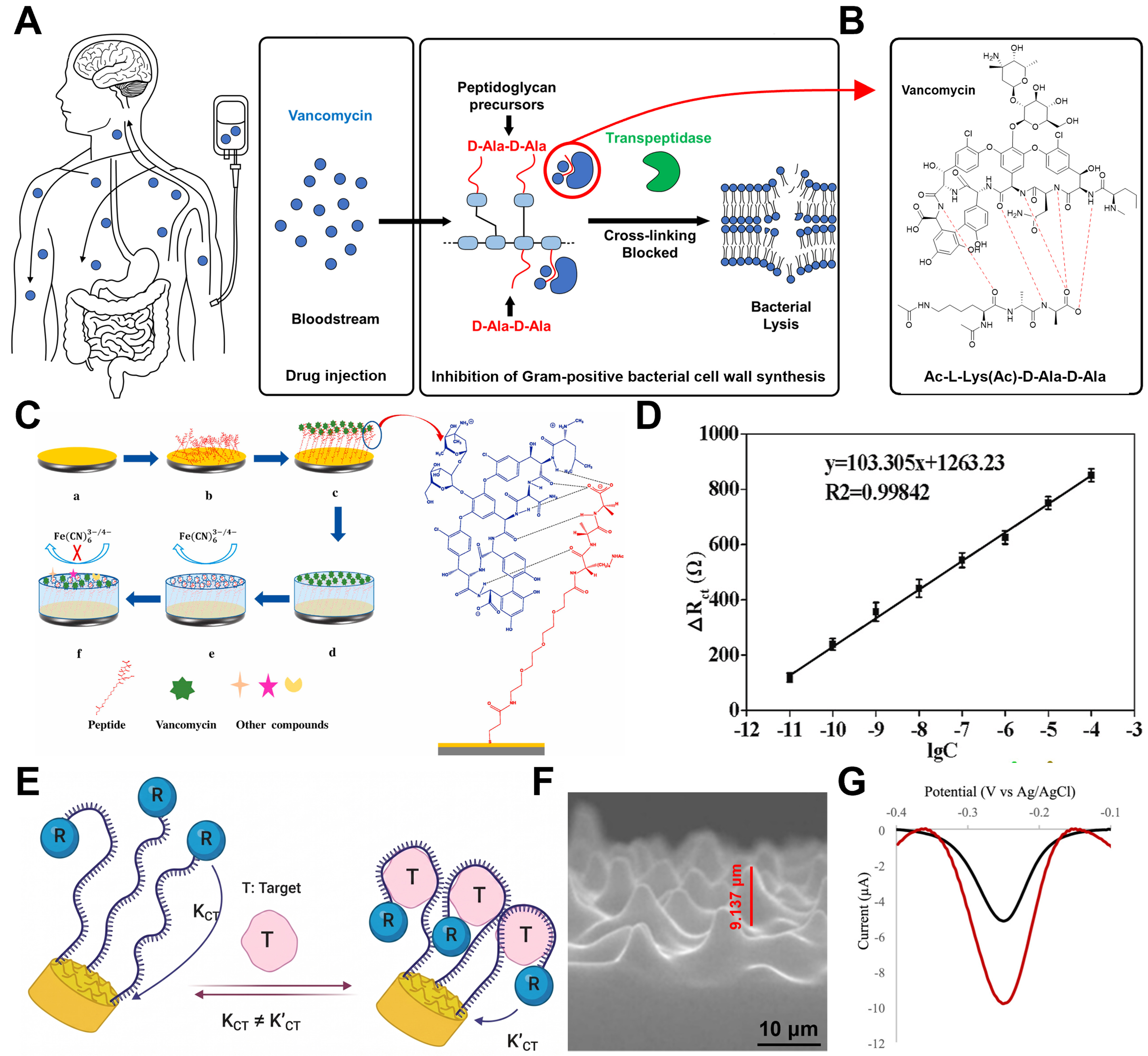
Figure 10. Vancomycin’s therapeutic mechanism for nephritis and sensing devices. (A) Bactericidal mechanism of vancomycin inhibiting cell wall synthesis by specifically binding to the D-Ala-D-Ala terminus; (B) Illustration of the five specific hydrogen bonds formed between vancomycin and the D-Ala-D-Ala terminus11; (C) Sensing principle of the hybrid peptide-MIP sensor, where target capture blocks electron transfer; (D) Linear calibration curve of electron transfer resistance ∆Rct) versus vancomycin concentration (R2 ≈ 0.998); (E) Schematic of the aptamer-based sensor using a 3D MSE for enhanced sensitivity; (F) Cross-sectional SEM image of the 3D MSE; (G) Comparison of SWV responses demonstrating the superior signal gain of the 3D MSE (red) over the planar electrode (black). The error bar in (D) represents the standard deviation. (C and D) Reproduced with permission Copyright 2021, Biosensors and Bioelectronics[134]. (E-G) Reproduced with permission Copyright 2025, ACS Omega[130]. R2: Coefficient of determination; 3D: three-dimensional; MSE: microstructured electrode; SEM: scanning electron microscope; SWV: square wave voltammetry.


