

Flexible and immunomodulatory milk-derived protein hydrogels as transient interfacing scaffolds for peripheral nerve integration
 0
0  ,
, Graphical Abstract
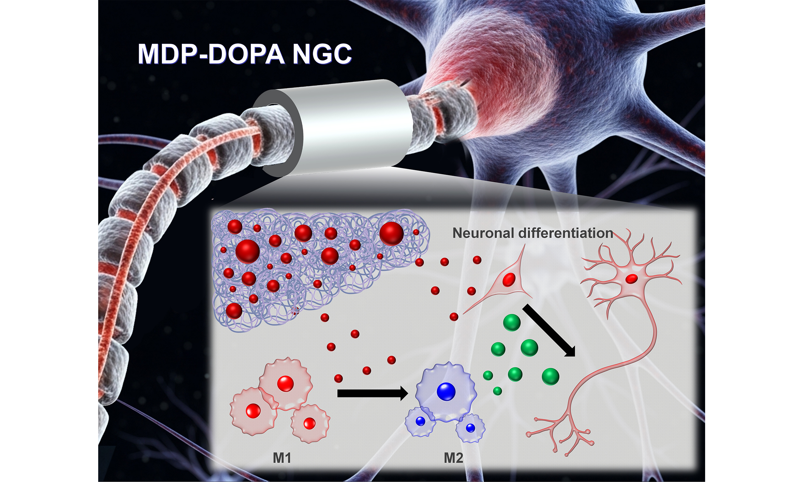
Abstract
Next-generation implantable neural interfaces require highly flexible, biocompatible, and transient material platforms to minimize mechanical mismatch with neural tissues and avoid secondary retrieval surgeries. This study developed a soft and biodegradable neural interfacing platform fabricated from milk-derived protein (MDP) and polyvinyl alcohol functionalized with 3,4-dihydroxyphenylalanine (DOPA) as an immunomodulatory nerve guidance conduit. The fabricated MDP-DOPA hydrogel conduits exhibited favorable protein-release kinetics, compliant mechanical stability that matched native neural tissues, and controlled biodegradation profiles. In vitro, the MDP-DOPA substrate significantly attenuated pro-inflammatory M1 macrophage polarization while selectively promoting the pro-regenerative M2 phenotype in RAW 264.7 cells, subsequently enhancing PC12 neuritogenesis through the bioactivity of released cascade peptides. In vivo, a rat 10-mm sciatic nerve defect model demonstrated that the implantation of soft MDP-DOPA conduits drastically improved structural and functional neural integration. This was evidenced by an enhanced sciatic functional index, elevated nerve conduction velocity, and robust axonal remyelination in the distal segments, closely approaching the recovery level of clinical gold-standard autografts. Furthermore, early-stage in vivo analysis verified that the platform successfully orchestrated a pro-regenerative immunomodulatory microenvironment at the interface site within 1 week. These findings highlight the potential of MDP-DOPA hydrogels as bioinstructive, soft, and stable electronic encapsulation or scaffolding platforms for next-generation implantable neural technologies for clinical translation.
Keywords


 Download PDF
INTRODUCTION
Annually, over 5 million people worldwide experience peripheral nerve injuries resulting from acute trauma, aging, and iatrogenic side effects, which can lead to loss of motor function, muscle paralysis, and lifelong disability[1,2]. Extensive efforts in recent decades have focused on developing engineered nerve guidance conduits (NGCs) that facilitate both structural and physiological restoration. Several NGCs have been approved by the Food and Drug Administration (FDA), such as Neurotube® and NeuraGen®, based on poly(glycolic acid) and collagen[3,4]. However, the effects of commercially available NGCs on nerve regeneration are insufficient to achieve physiological functional recovery, including sensation and motility, comparable to those of autografts[5]. Functional NGCs facilitate inflammation, proliferation, and remodeling during nerve regeneration[6]. During the inflammatory phase, a sequential chain of molecular and cellular events known as Wallerian degeneration occurs. M1 macrophages, which respond to pro-inflammatory cytokines, promote the clearance of debris and angiogenesis and then switch to M2 macrophages, which stimulate the next phase of nerve regeneration: the proliferation and migration of Schwann cells that form an aligned Schwann cell cable. In the remodeling phase, the recruited Schwann cells lead to axonal sprouting from the proximal to the distal region of the defective nerve gap using a guidance mechanism[7-9].
Several studies have developed functional NGCs using additional biological stimuli, including exogenous drugs or growth factors such as interleukin 10 (IL-10) and nerve growth factor (NGF). These factors accelerate inflammation and recruit various cells to induce remodeling[10,11]. Although these additional growth factors have proven therapeutic effects, their use as therapeutic agents is limited because of their short half-life, side effects at high doses, and loss of activity when loaded onto various types of NGCs[12]. Therefore, we considered applying appropriate bioactive biomaterials to accelerate nerve regeneration. Milk-derived protein (MDP) casein is a suitable biomaterial base for releasing various bioactive peptides through proteolytic enzymes and hydrolysis under normal body conditions[13]. A key advantage of MDP is its immunomodulatory effects, driven by bioactive peptides such as opioid peptides, β-casomorphins, β-casochemotide, κ-casein, and FLPYPY[14-17]. Immunopeptides stimulate phagocytosis by recruiting macrophages in vivo. Several bioactive casein peptides affect each phase of nerve regeneration. In the inflammation phase, β-casochemotide-1 and κ-casein recruit macrophages via chemotaxis and induce an M2-like phenotype by reducing toll-like receptor (TLR)-induced cytokine production[15,16]. In addition, β-casomorphins and FLPYPY-containing peptides can stimulate neurite outgrowth[14,17]. Thus, we fabricated MDP-NGCs to investigate the effect of bioactive peptides on MDP and to help orchestrate peripheral nerve regeneration steps.
Although casein shows great potential as a tissue-engineering biomaterial, it has limited mechanical properties and may lack long-term structural stability because it is a natural polymer[18]. Polyvinyl alcohol (PVA), an FDA-approved biocompatible synthetic polymer with excellent mechanical properties, was used to address these limitations[19]. PVA can thermally crystallize the hydrogel phase through repeated simple freeze–thaw cycles without any chemical crosslinking agents[20]. A highly hydrophilic PVA hydrogel causes the reversible adsorption of proteins[21]. We immobilized 3,4-dihydroxyphenylalanine (DOPA), which is derived from mussel adhesive proteins, onto a hydrogel (MDP-DOPA) to investigate the bioactivity of the physical adsorption of bioactive molecules released from MDP[22]. In our previous study, the MDP-PVA scaffold showed a good protein release profile and function, including macrophage and mesenchymal stem cell recruitment[23].
In this study, we hypothesized that bioactive peptides released from MDP-DOPA could accelerate nerve regeneration by regulating immune responses and neurite outgrowth. To test our hypothesis, we cast a blended MDP and PVA solution in a cylindrical mold and used a simple freeze–thaw cycle to fabricate a flexible NGC (MDP) and immobilized DOPA (MDP-DOPA) for implantation into defective sciatic nerves. Morphological, chemical, and mechanical analyses were performed on the fabricated flexible MDP and MDP-DOPA hydrogel conduits. A polarization study using RAW 264.7 cells, cultured on each hydrogel surface, was performed to confirm M1/M2 polarization. The neurogenic differentiation of PC12 cells was investigated using conditioned media (CM) containing various cytokines from RAW 264.7 cells cultured on MDP and MDP-DOPA hydrogels. Finally, each NGC was implanted in a 10-mm rat sciatic nerve defect, and an autograft (the clinical gold standard) was used as the positive control. We investigated the initial reaction and regeneration of the defective nerve using functional analyses such as the sciatic function index (SFI), electrophysiological tests, and muscle atrophy, as well as histological analysis using immunohistochemistry (IHC) and osmium tetroxide staining.
EXPERIMENTAL
Preparation of MDP-DOPA hydrogels
Following the physical crosslinking protocols adapted from our earlier work[23], physical crosslinking of the MDP-DOPA network was achieved through repeated freeze–thaw thermal cycling. Briefly, bovine casein (5% w/v; C5890, Sigma-Aldrich, St. Louis, MO, USA) was dissolved in 1 M NaOH, whereas PVA (4% w/v) was solubilized in distilled water (DW) at 90 °C. Before blending, the hot PVA solution was cooled to RT to prevent the thermal denaturation of the protein. The two solutions were mixed thoroughly overnight under continuous stirring at room temperature. The mixed solutions were poured into various casting molds (flat and conduit forms). For in vitro bioevaluations and cellular assays, planar substrates were fabricated using a mold consisting of dual parallel polystyrene sheets separated by a 1.5-mm spacer. The conduit mold consisted of an outer conduit and an inner glass tube to analyze its mechanical properties and conduct in vivo animal studies. The final hydrated conduit had an inner diameter of 1.11 ± 0.08 mm and a wall thickness of 0.80 ± 0.04 mm [mean ± standard deviation (SD), n = 5]. After casting the mixed solution, the molds underwent freeze–thaw cycles. To induce hydrogel crosslinking, the loaded molds underwent five iterative freeze–thaw steps, comprising deep-freezing at -80 °C for 4 h and subsequent ambient thawing for 2 h. The fabricated hydrogel was then washed to remove undesired reactants and byproducts. MDP was modified with DOPA (D9628; Sigma-Aldrich, St. Louis, MO, USA) to immobilize biomolecules and improve cell adhesion. Surface modification was performed by incubating the MDP hydrogels in a 10 mM Tris–HCl solution (pH 8.5; T1503, Sigma-Aldrich, St. Louis, MO, USA) supplemented with 2 mg·mL-1 DOPA for 16 h at RT. Under these alkaline conditions, the DOPA monomers underwent self-oxidation and polymerization to form a stable poly-DOPA-like coating on the hydrogel surface. After coating, the specimens were thoroughly sonicated in DW to dislodge and remove loosely bound poly-DOPA aggregates or nonspecifically adsorbed particulates. This rigorous cleaning step ensured that only firmly immobilized and chemically cross-linked polymeric layers remained on the substrate.
Morphological analysis of MDP-DOPA substrate
The external and internal surfaces of the MDP and MDP-DOPA NGCs were mounted on an aluminum stub. To prepare for the scanning electron microscopy (SEM) observations, the NGC samples were sputter-coated with platinum using an automated system (Sputter Coater 108 Auto, Cressington Scientific Instruments, Watford, UK) under vacuum conditions, operating at a 15 mA plasma current for 2 min. The coated samples were characterized by SEM (MERLIN Gemini 2, Carl Zeiss, Oberkochen, Germany; facility equipment registration no. NFEC-2021-05-270500) at the Center for Biomedical Engineering Core Facility (Dankook University, Cheonan, Republic of Korea), at an accelerating voltage of 10 kV.
Evaluation of the permeability and mechanical properties of MDP-DOPA NGCs
Hydrogel permeability was evaluated by monitoring the diffusion of toluidine blue (TB; 100 μM; T3260, Sigma-Aldrich, St. Louis, MO, USA) across planar hydrogel disks (n = 5) with a multimode microplate reader (Spark 20M, Tecan, Männedorf, Switzerland). We prepared flat disc-type hydrogels (thickness =
Degradation profile of MDP-DOPA NGCs
To evaluate enzymatic degradation profiles, MDP-DOPA hydrogel disks (20 mm in diameter, n = 5) were placed in 5 mL of a 1 mg·mL-1 collagenase B solution (11088815001, Roche Diagnostics, Basel, Switzerland) prepared in phosphate-buffered saline (PBS, pH 7.4; P3813, Sigma-Aldrich, St. Louis, MO, USA) and maintained at 37 °C with gentle agitation at 30 rpm[24,25]. Unmodified MDP hydrogel disks were processed alongside MDP-DOPA samples as comparative controls under identical conditions, with n = 5 per experimental group. Each hydrogel was weighed at different time points (1, 2, 4, 6, and 8 weeks) after washing with DW and complete lyophilization to remove residual water. Day 1 was included solely to show the initial appearance of the samples after fabrication and before initiation of the degradation test. The weight loss at each time point (t) was calculated using the following equation: weight loss (%) = (Wi - Wt)/Wi × 100, where Wi represents the initial weight of the hydrogel at day 0, and Wt represents the weight of the hydrogel at each time point.
MDP-DOPA CM collection and M1/M2 macrophage polarization test
RAW 264.7 macrophages (RRID: CVCL_0493; ATCC, Manassas, VA, USA, as certified by the vendor) were cultured on planar MDP and MDP-DOPA hydrogel substrates measuring 100 mm in diameter (1 × 10⁶ cells hydrogel-1, n = 4) with lipopolysaccharide (LPS; 250 ng·mL-1, L2630, Sigma-Aldrich, St. Louis, MO, USA)-treated media [PM; 1% penicillin/streptomycin (PS; 30-002-CI, Corning, Corning, NY, USA) and 10% fetal bovine serum (FBS; 35-010-CV, Corning, Corning, NY, USA) in Dulbecco’s modified eagle medium (DMEM; 10-013-CV, Corning, Corning, NY, USA)] for 24 h to evaluate the ratio of M1/M2 macrophage polarization. The cells were then washed three times with PBS and treated with the proliferation medium. The CM were collected from RAW 264.7 cells incubated with tissue culture polystyrene (TCPS, 430167, Corning, Corning, NY, USA), C-CM, MDP (M-CM), and MDP-DOPA (MD-CM), centrifuged for 20 min at 300 g, and filtered with a 0.45-μm syringe filter (431220, Corning, Corning, NY, USA). RAW 264.7 cells were detached from each hydrogel, and M1/M2-related genes were evaluated. Total cellular RNA was isolated using an AccuPrep® Universal RNA Extraction Kit (K-3140, Bioneer, Daejeon, Republic of Korea) according to the manufacturer’s protocol. RNA concentration and optical purity ratios (A260/A280) were assessed spectrophotometrically using a NanoDrop One instrument (Thermo Fisher Scientific, Waltham, MA, USA). For cDNA synthesis, 1 μg of the isolated RNA template was reverse-transcribed into complementary DNA utilizing AccuPower® CycleScript RT PreMix (K-2044, Bioneer). The resulting cDNA served as a template to quantify M1/M2 macrophage-specific gene target levels via real-time polymerase chain reaction (PCR) using SYBR Green. Target transcription was determined using the comparative 2-ΔΔCT approach, with results expressed as relative fold-change compared to RAW 264.7 cells cultivated on standard TCPS. Other surface markers of RAW 264.7 cells were analyzed using the intraclass correlation coefficient. To fix the cultured RAW 264.7 cells, specimens were incubated in 4% paraformaldehyde (PFA; 58127, Sigma-Aldrich, St. Louis, MO, USA) for 15 min and subsequently washed three times with PBS. To prevent non-specific binding, the washed cells were incubated in a blocking solution consisting of 5% goat serum (G9023, Sigma-Aldrich, St. Louis, MO, USA) and 0.1% Triton™ X-100 (T8787, Sigma-Aldrich) in PBS for 1 h at ambient temperature. The samples were then probed with primary antibodies against CD206 (PA5-101657, Invitrogen, Thermo Fisher Scientific, Carlsbad, CA, USA) and CD86 (ab220188, Abcam, Cambridge, UK) overnight at 4 °C. For signal visualization, the secondary antibodies were conjugated to fluorescein isothiocyanate (FITC; A-11012, Invitrogen) and rhodamine (A-11008, Invitrogen), followed by nuclear counterstaining with 4′,6-diamidino-2-phenylindole (DAPI; Vectashield, H-1200, Vector Laboratories, Burlingame, CA, USA). Fluorescence imaging was performed using a fluorescence microscope (Eclipse Ts2R; Nikon, Tokyo, Japan), and target-positive signals were quantified using ImageJ software (NIH, Bethesda, MD, USA).
Examination of neurogenic differentiation using CM
To investigate the synergistic relationship between the retrieved CM and neurogenic cues, each CM was thoroughly mixed with neuronal differentiation media (NDM; DMEM supplemented with 10% FBS, 5% horse serum (H1138, Sigma-Aldrich, St. Louis, MO, USA), 1% PS, and NGF at 100 ng·mL-1 at a volumetric ratio of 6.5:3.5 (CM:NDM), yielding a sub-optimal final NGF concentration of 35 ng·mL-1 within the culture microenvironment. After culturing 5 × 10³ PC12 [RRID: CVCL_0481, American Type Culture Collection (ATCC), Manassas, VA, USA] cells in each well of a 96-well plate (3599, Corning, Corning, NY, USA) in various CMs, the cells were observed under a light microscope (AZ100, Nikon, Tokyo, Japan) at each time point (biological n = 4). Morphometric quantification, including neurite length and the proportion of neurite-positive cells (defined as cells exhibiting processes exceeding 10 μm), was performed on captured images using ImageJ.
Implantation of MDP and MDP-DOPA NGCs in rat sciatic nerve defects
Adult male SD rats (8 weeks old, weight: 260 ± 20 g at the time of surgery; Dayun, Gyeonggi-do, Republic of Korea) were used to make the sciatic nerve defect model, as previously described[26]. All experimental procedures involving animals were approved by the Institutional Animal Care and Use Committee of Dankook University (approval no. DKU-22-001) and complied strictly with the NIH Guide for the Care and Use of Laboratory Animals. Animals were maintained in individual cages under controlled environmental conditions (a temperature of 23-25 °C and 45%-50% relative humidity) with ad libitum access to food and water. After 1 week of housing, surgical interventions were performed under general anesthesia induced with isoflurane (Forane®, Choongwae Pharma, Seoul, Republic of Korea). After hair removal, an incision was made through the skin and underlying muscle of the left gluteal region to expose the sciatic nerve. A 10-mm nerve defect was created by completely transecting and excising the nerve 5 mm distal to the hip joint. Both the proximal and distal nerve stumps were subsequently inserted into a 14-mm-long NGC (n = 4 per group) and coapted into the epineurium using 10-0 nylon sutures (NK 1013; AILEE Co., Busan, Republic of Korea). At each evaluation time point (1, 4, and 8 weeks post-implantation), 4 animals per group were assigned. Finally, the muscular, subcutaneous, and cutaneous layers were sequentially closed using 6-0 (SK 617; AILEE Co.) and 3-0 (SK 312; AILEE Co.) silk sutures. All rats were euthanized using CO2 at weeks 1, 4, and 8, and their hind limbs were retrieved. All in vitro experiments were conducted between February and December 2021, and all in vivo experiments were conducted between February 2022 and 2023.
Sciatic nerve functional recovery evaluation
Axonal functional recovery post-implantation was determined using gastrocnemius electromyography (EMG) recordings and SFI measurements. For EMG evaluation, the rats were anesthetized using isoflurane, and electrical stimulation (1.5 mA) and signal recording were performed using the Medelec Synergy system (Oxford Instruments Medical Inc., Surrey, UK). To determine SFI values, footprints were collected biweekly by having the rats traverse a custom walking track lined with white paper. The print length, toe spread, and intermediate toe spread were measured and applied using the following formula: SFI = -38.3 × [(EPL - NPL) NPL-1] + 109.5 × [(ETS - NTS) NTS-1] - 13.3 × [(EIT - NIT) NIT-1] - 8.8, where EPL is the experimental print length, NPL is the normal print length, ETS is the experimental toe spread, NTS is the normal toe spread, EIT is the experimental intermediary toe spread, and NIT is the normal intermediary toe spread.
Investigation of muscle atrophy and histological analysis
To assess nerve injury-induced muscle atrophy, the gastrocnemius muscles were harvested at 4 and 8 weeks post-implantation. After rinsing with PBS, wet weights of the harvested muscles were immediately recorded. For tissue preparation, samples were fixed overnight in 4% PFA, dehydrated through a graded sucrose gradient (10%, 15%, and 30%) for 2 h each at 4 °C, and embedded in OCT compound (4583, Sakura Finetek USA Inc., Torrance, CA, USA) prior to cryosectioning. The sections were stained with hematoxylin (GHS232, Sigma-Aldrich, St. Louis, MO, USA) and eosin (HT110232, Sigma-Aldrich, St. Louis, MO, USA) [hematoxylin and eosin (H&E)] and observed by light microscopy. Muscle fiber diameters were quantified from the captured histological images using the ImageJ software (National Institutes of Health, Bethesda, MD, USA).
Immunohistochemical and quantitative reverse transcription polymerase chain reaction analyses
The sciatic nerve of each group was harvested 1 week after implantation. The harvested nerves were snap-frozen and divided into the proximal region and remaining tissue. The proximal regions were used for IHC analysis, and the remaining regions were used for quantitative reverse transcription polymerase chain reaction (qRT-PCR) analysis. The RNA was isolated according to the manufacturer’s instructions. Other processes were the same as those used in the in vitro qRT-PCR analysis, with the autograft group serving as the reference calibrator (relative expression level = 1) for the 2-ΔΔCt calculation. Four and eight weeks after implantation, the harvested nerves were subjected to IHC and osmic acid staining. For IHC analysis, the harvested nerve tissues were fixed in 4% PFA and cryo-sectioned via a Leica CM3050 S cryostat (Leica Microsystems, Wetzlar, Germany). Permeabilization and blocking of nonspecific binding were performed using 0.3% Triton X-100 supplemented with 5% goat serum for 1 h at room temperature. The prepared sections were then incubated with the following target primary antibodies for 1-week specimens: rabbit anti-CD86 (1:100; Invitrogen, Thermo Fisher Scientific, Carlsbad, CA, USA) and rabbit anti-CD206 (1:100; Bioss, Woburn, MA, USA). For 4- and 8-week specimens, rabbit anti-neurofilament 200 monoclonal (1:300; Abcam, Cambridge, UK) and rabbit anti-S-100 polyclonal antibodies (1:1,000; Abcam) were used. Following three sequential PBS washes, tissue sections were incubated for 1 h at room temperature with fluorophore-conjugated secondary antibodies, either FITC- or rhodamine-labeled goat anti-rabbit IgG (1:200; Jackson ImmunoResearch Laboratories, West Grove, PA, USA), diluted in PBS containing 1% goat serum. Nuclear counterstaining was performed using DAPI. Immuno-stained specimens were photographed using a fluorescence microscope. All positive signals for each antibody were evaluated using the ImageJ software. For osmic acid staining, the samples were fixed with 4% PFA and stained with osmic acid (1% w/v) (75632, Sigma-Aldrich, St. Louis, MO, USA). After washing with excess DW for 6 h, paraffin sections (3 µm) were cut and observed by light microscopy. The thickness of the myelin sheath and axon diameter were calculated using the ImageJ software.
Statistical analysis
Quantitative data are expressed as means ± SDs. The SPSS software (IBM SPSS Statistics) was used to perform Bonferroni and Tukey tests.
RESULTS AND DISCUSSION
Results
Fabrication and morphological characterization of MDP and MDP-DOPA NGCs
The MDP and PVA solutions were mixed, cast in a conduit mold, and physically crosslinked through five freeze–thaw cycles. This process induced physical crosslinking between PVA and MDP, resulting in a phase transition to form a hydrogel without chemical crosslinkers [Figure 1A][20]. The MDP-DOPA NGC was designed to provide sustained release of MDP-derived bioactive peptides to modulate the regenerative microenvironment through M2 macrophage polarization and enhanced expression of regenerative factors [Figure 1B]. The fabricated MDP NGC was then coated with DOPA[27] to enhance the immobilization of bioactive molecules on its surface. To monitor the MDP release profile, cumulative protein release from the hydrogels was quantified over 28 days [Supplementary Figure 1]. A comparison between MDP and MDP-DOPA demonstrated a highly stable, sustained release profile, gradually liberating approximately 25% of the total protein by day 28 without any noticeable initial burst release. Macroscopically, both uncoated (MDP) and DOPA-coated (MDP-DOPA) NGCs were morphologically similar, except for their color, with MDP-DOPA being darker [Figure 1C]. Both conduits had an inner diameter of 1.11 ± 0.08 mm and a wall thickness of 0.80 ± 0.04 mm. SEM images of the surface and internal cross sections revealed that both MDP and MDP-DOPA possessed highly porous structures formed by ice crystals during freezing, with no significant morphological differences observed between the two groups. The apparent lamellar morphology observed in the lyophilized samples resulted from ice crystal growth and phase separation during the freeze–thaw process and subsequent lyophilization, rather than representing the native architecture of the hydrated hydrogels. The microstructural analysis revealed a heterogeneous pore distribution with an average pore diameter of 5.18 ± 3.21 μm, spanning from approximately 1 to 10 μm as systematically detailed in the statistical histogram [Figure 1D and Supplementary Figure 2][26].
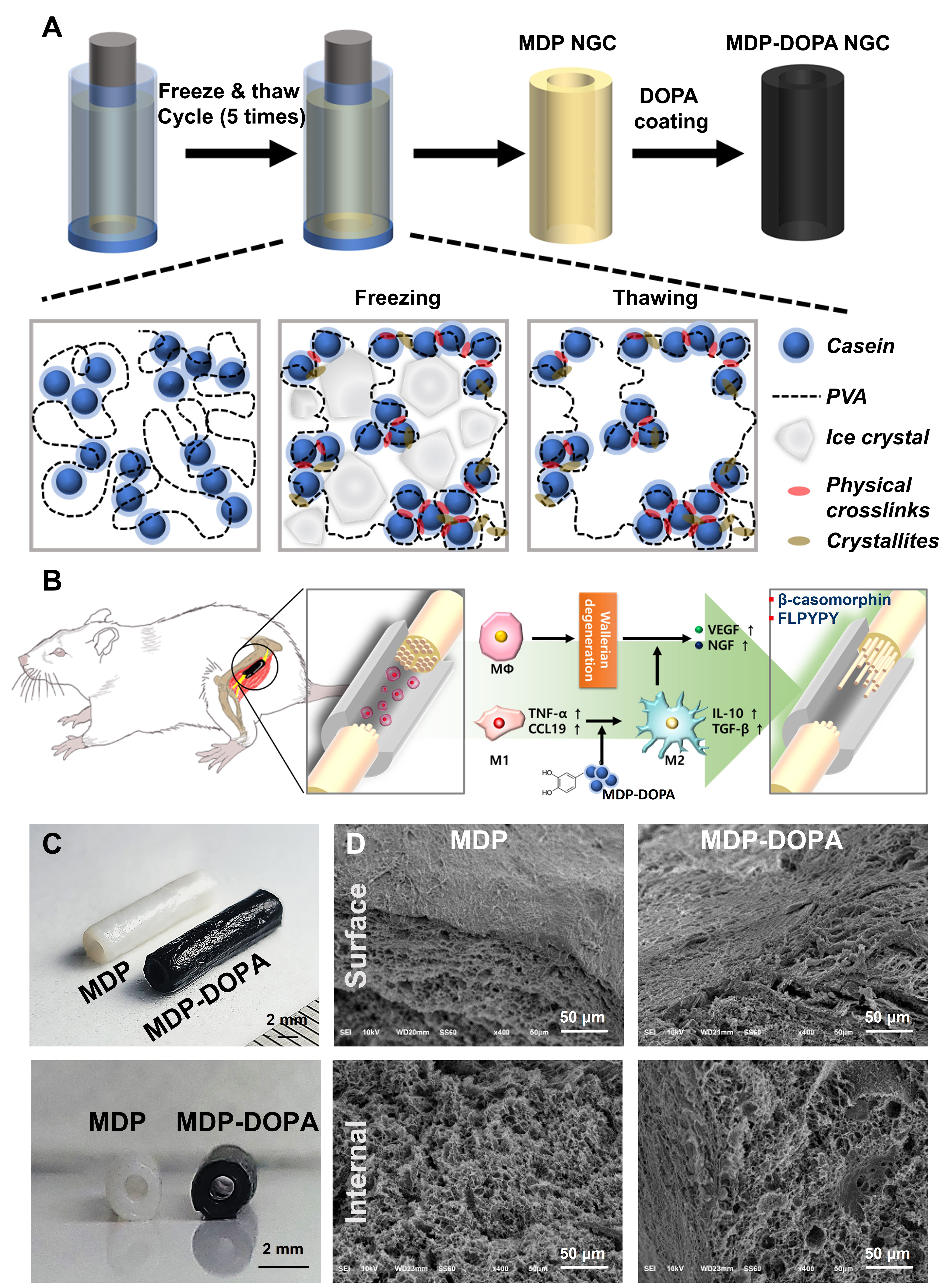
Figure 1. (A) Fabrication process of the MDP-DOPA hydrogel conduit and (B) treatment strategy using MDP-DOPA for nerve regeneration; (C) Each fabricated NGC; (D) Fabricated MDP and MDP-DOPA NGCs (Scale bar = 50 μm). MDP-DOPA: Milk-derived protein-dihydroxyphenylalanine; NGC: nerve guidance conduit; PVA: polyvinyl alcohol; VEGF: vascular endothelial growth factor; NGF: nerve growth factor.
Physicochemical and mechanical properties of MDP-DOPA NGCs
The permeability of NGCs was assessed using the TB diffusion test [Figure 2A]. After 180 min, the concentration of diffused TB was 5.63 ± 0.15 µM for MDP and 5.07 ± 0.18 µM for MDP-DOPA, with no statistically significant variance between the two groups [Figure 2A][28,29]. Compressive and tensile strength analyses were performed to characterize the mechanical behavior of the conduits [Figure 2B and C]. The compressive elastic modulus of MDP (1,025.30 ± 36.40 kPa) and MDP-DOPA (956.20 ± 56.44 kPa) were not significantly different [Figure 2B]. Under tensile loading, both hydrogel formulations demonstrated remarkable elongation capacity, yielding comparable Young’s modulus values (0.10 ± 0.001 MPa for MDP vs. 0.09 ± 0.01 MPa for MDP-DOPA) [Figure 2C].
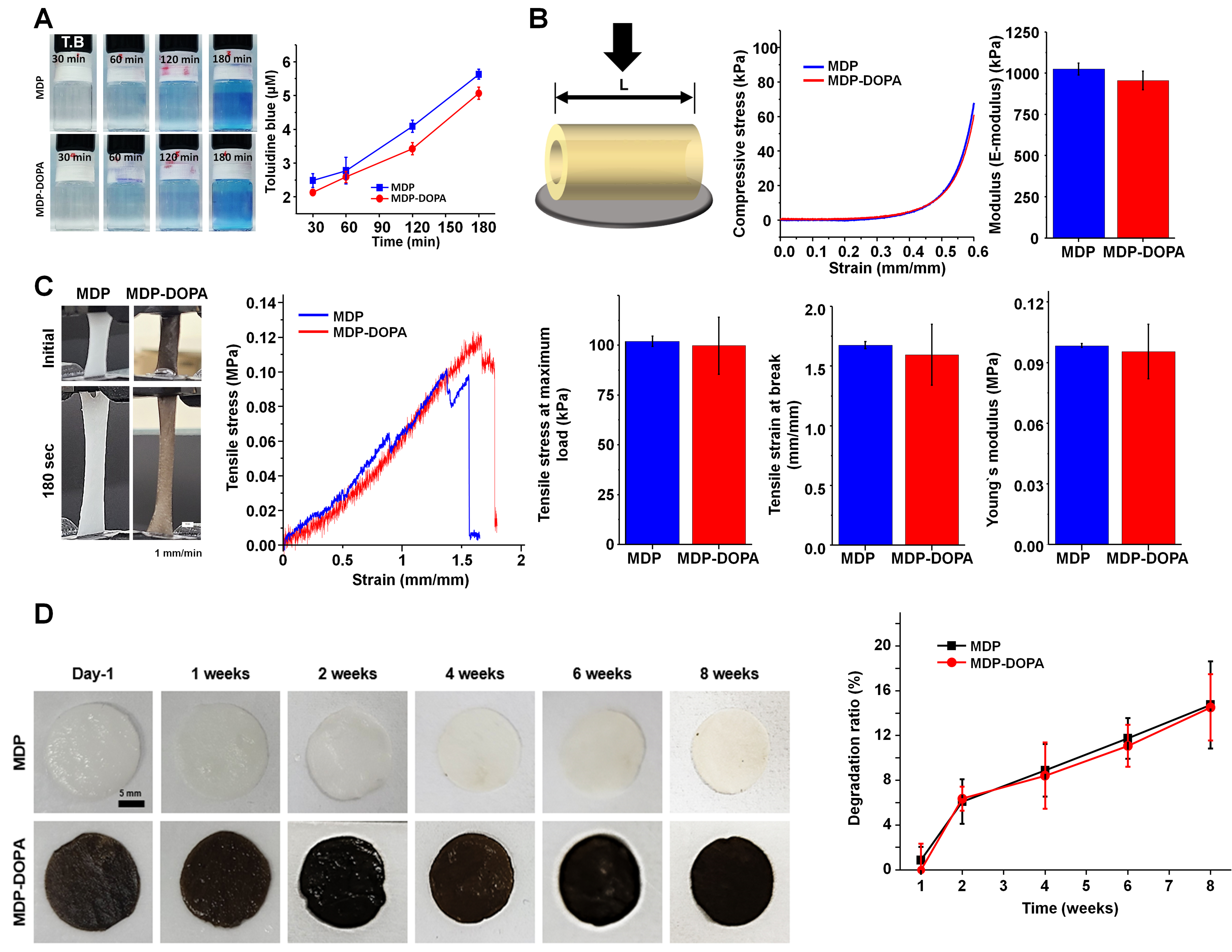
Figure 2. Characterization of MDP and MDP-DOPA. (A) In vitro permeability test of MDP and MDP-DOPA using TB solution (n = 5); Evaluation of mechanical properties: (B) compressive modulus (n = 5, crosshead speed: 1 mm·min-1) and (C) tensile properties (n = 5, crosshead speed: 1 mm·min-1); (D) In vitro degradation test of MDP and MDP-DOPA. Representative images of degraded hydrogels at each time point (n = 5, scale bar = 5 mm). All values are reported as mean ± SD from five independent hydrogel specimens per group. MDP: Milk-derived protein; DOPA: dihydroxyphenylalanine; TB: toluidine blue; SD: standard deviation.
An in vitro degradation test was performed over 8 weeks [Figure 2D]. After 8 weeks, MDP and MDP-DOPA showed similar degradation rates, maintaining their scaffolding ability throughout the test. Altogether, these findings demonstrate that surface functionalization with DOPA preserved both the microstructural architecture and mechanical integrity of the hydrogel matrix[30,31]. Furthermore, the mechanical properties obtained in this study are considered suitable for peripheral NGCs because nerve repair is typically performed under tension-free coaptation and does not require high tensile loading[32]. Consistently, the implanted conduits maintained their tubular architecture without any apparent lumen collapse throughout the experimental period, as confirmed by fluorescence imaging [Supplementary Figure 3].
MDP-DOPA promoted M2 macrophage polarization and neurite outgrowth in vitro
To investigate the immunomodulatory effects, the expression of M1 (TNF-α, CD86, CCL19, and CXCL11) and M2 (IL-10, CD163, CCL13, and CD206) macrophage markers was investigated in LPS-activated RAW 264.7 cells that were cultured on TCPS, MDP, or MDP-DOPA substrates [Figure 3A and B]. The expression of M1 markers generally decreased in the MDP-DOPA group, whereas M2 markers were significantly increased in both the MDP-treated- and MDP-DOPA-treated groups compared with the control [Figure 3B]. To verify whether M2 polarization was independently induced by DOPA or the MDP matrix, polycaprolactone and DOPA-coated polycaprolactone were evaluated as non-bioactive control substrates. As presented in Supplementary Figure 4, the absence of M2 marker upregulation on DOPA-coated polycaprolactone confirmed that the immunomodulatory M2 polarization is primarily driven by the bioactive MDP matrix rather than the DOPA coating alone. These results were validated via immunofluorescence, which revealed downregulation of the pro-inflammatory marker CD86 alongside robust CD206 expression in macrophages grown on the MDP-DOPA substrate [Figure 3C and D][6,7,33]. To further corroborate this phenotypic transition at the protein secretion level, a comprehensive cytokine profiling of the macrophage secretome was performed using a protein array assay [Supplementary Figure 5]. Relative to the unmodified MDP substrate, the MDP-DOPA hydrogel strongly suppressed key pro-inflammatory M1 mediators, including TNF-α, IL-1β, IL-6, IL-12 p40, CCL2, and CXCL10. Concurrently, dramatic upregulation was observed in anti-inflammatory M2 markers (IL-10, IL-13, and Chitinase 3), as well as in key neurotrophic, angiogenic, and cell-survival growth factors, including vascular endothelial growth factor (VEGF), HGF, EGF, LIF, and Gas6.
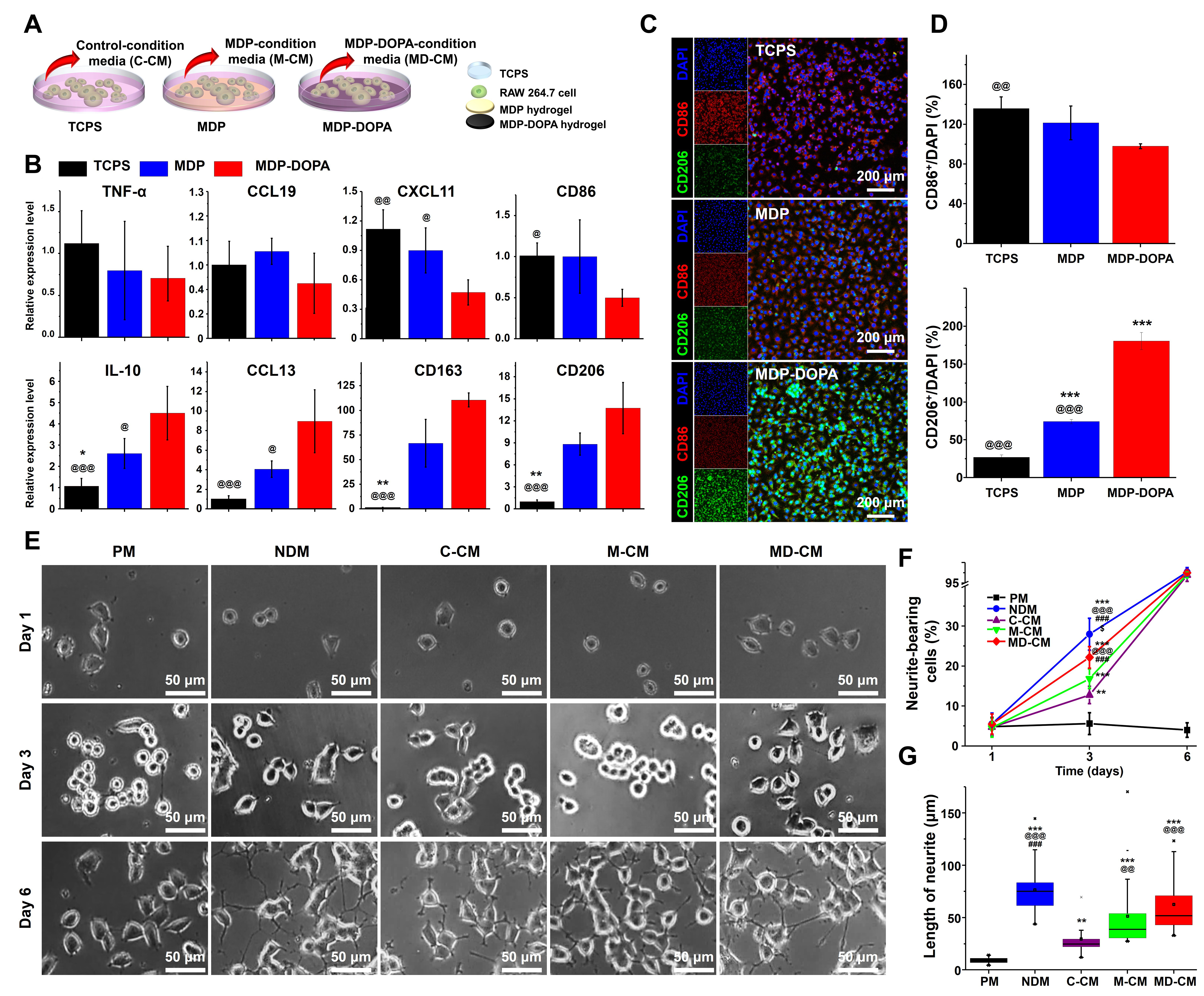
Figure 3. (A) Schematic illustration of collecting various CM; (B) Examination of M1/M2 macrophage phenotype-related markers. (*P < 0.05 and **P < 0.01 vs. MDP; @P < 0.05,@@P < 0.01, and @@@P < 0.001 vs. MDP-DOPA); (C) Confocal micrographs and (D) corresponding quantitative analysis of M1 (CD86) and M2 (CD206) phenotypic markers in cultured macrophages. (***P < 0.001 vs. TCPS, @@P < 0.01 and @@@P < 0.001 vs. MDP-DOPA), (Scale bar = 200 μm); (E) Representative phase-contrast microscopic images showing differentiated PC12 cells cultured in various CM (Scale bar = 50 μm); (F) Proportion of cells with neurites, and (G) neurite lengths for each sample (**P < 0.01 and ***P < 0.001 vs. PM, @@P < 0.01 and @@@P < 0.001 vs. C-CM, ###P < 0.001 vs. M-CM, $P < 0.05 vs. MD-CM). In (G), data are presented as a box-and-whisker plot, where the center line represents the median, the box boundaries indicate the 25th and 75th percentiles (IQR), the whiskers denote 1.5 × IQR, and individual outlier points are shown as markers. For all other quantitative panels (B, D, and F), values represent the mean ± SD derived from four biological replicates (n = 4). CM: Conditioned media; MDP: milk-derived protein; DOPA: dihydroxyphenylalanine; TCPS: tissue culture polystyrene; PM: proliferation media; C-CM: TCPS conditioned media; M-CM: MDP conditioned media; MD-CM: MDP-DOPA conditioned media; IQR: interquartile range; SD: standard deviation; DAPI: 4′,6-diamidino-2-phenylindole; NDM: neuronal differentiation media.
Next, the effect of CM from these macrophage cultures on neurite outgrowth was tested using PC12 cells [Figure 3E]. By day 3, the proportion of cells exhibiting neurite outgrowth reached a significantly higher level within the NDM (28.00% ± 3.99%) and MDP-DOPA conditioned media (MD-CM, 22.20% ± 2.71%) groups compared to the proliferation media (PM, 5.60% ± 2.74%), TCPS conditioned media (C-CM, 12.77% ± 2.18%), and MDP conditioned media (M-CM, 16.84% ± 2.42%) groups [Figure 3F]. Similarly, neurite length at day 3 was significantly longer in the NDM (76.4 ± 20.7 μm), MD-CM (62.4 ± 26.0 μm), and M-CM (51.3 ± 33.0 μm) groups than in the PM (9.0 ± 2.8 μm) and C-CM (29.5 ±
MDP-DOPA NGCs enhance functional recovery of injured sciatic nerve
To assess the regenerative performance of the conduits, a 10-mm rat sciatic nerve defect model was established, with autografts serving as a positive control [Figure 4A][26]. SFI values were determined to evaluate motor function recovery. At 8 weeks post-implantation, SFI values in the autograft (-65.35 ± 2.12) and MDP-DOPA (-71.48 ± 0.63) groups exhibited statistically superior levels relative to the MDP group (-80.19 ± 1.53) [Figure 4B and C].
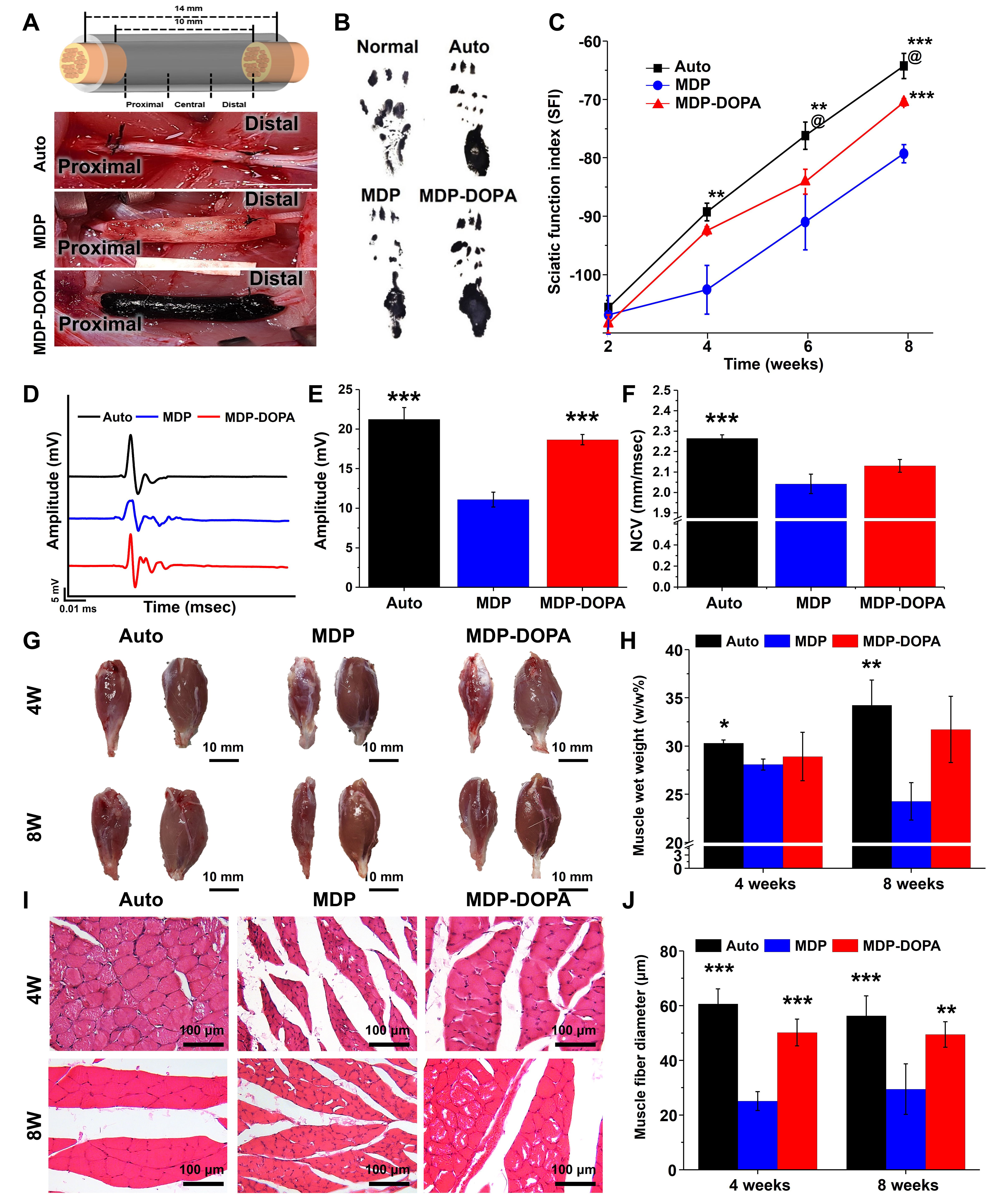
Figure 4. (A) Schematic illustration of NGC implantation and macroscopic images of implanted NGCs; (B) Paw printing at 8 weeks post-implantation and (C) quantification of sciatic functional index (**P < 0.05 and ***P < 0.001 vs. MDP, @P < 0.05 vs. MDP-DOPA); (D) Electrophysiological evaluation of compound muscle action potentials, (E) peak amplitude measurements, and (F) nerve conduction velocity of each group (***P < 0.001 vs. MDP); (G) Gross view of gastrocnemius muscle (scale bar = 10 mm) and (H) quantification of muscle wet weight ratio; (I) H&E histological sections of isolated gastrocnemius muscle (Scale bar = 100 μm) and (J) quantification of muscle fiber diameter. (*P < 0.05, **P < 0.01, and ***P < 0.001 vs. MDP). Data are presented as mean ± SD (biological n = 4). NGC: Nerve guidance conduit; MDP: milk-derived protein; DOPA: dihydroxyphenylalanine; H&E: hematoxylin and eosin; SD: standard deviation.
EMG at 8 weeks revealed that onset-to-peak amplitudes in the autograft (21.24 ± 1.50 mV) and MDP-DOPA (18.64 ± 0.67 mV) groups were significantly higher than in the MDP group (11.09 ± 0.95 mV) [Figure 4D and E]. NCV was also higher in the MDP-DOPA (2.13 ± 0.03 mm·ms-1) and autograft (2.26 ± 0.01 mm·ms-1) groups than in the MDP group (2.04 ± 0.05 mm·ms-1) [Figure 4F].
Muscle atrophy prevention was assessed by measuring the wet weight of the gastrocnemius muscle [Figure 4G and H][37,38]. At 8 weeks, the relative muscle wet weight was maintained in the autograft (34.24% ± 2.60%) and MDP-DOPA (31.72% ± 3.44%) groups, whereas it decreased further in the MDP group (24.26% ± 1.95%) compared to the 4-week time point. Histological analysis with H&E staining showed that at 8 weeks, muscle fiber diameter was significantly greater in the autograft (56.27 ± 7.35 µm) and MDP-DOPA (49.46 ± 4.67 µm) groups than in the MDP group (29.50 ± 9.26 µm) [Figure 4I and J].
Histological and morphometric analysis of nerve regeneration
Histological analyses of the regenerated nerves were performed at 4 and 8 weeks. Immunostaining for neurofilament 200 kDa (NF200) was performed to assess axonal outgrowth [Figure 5A]. At 8 weeks, the percentage of NF200-positive area in the central (28.33% ± 3.05%) and distal (14.06% ± 0.89%) regions demonstrated a marked elevation in the MDP-DOPA compared to the MDP (central; 19.15% ± 0.84%, distal; 3.22% ± 1.20%) and was similar to the autograft group [Figure 5B]. S100 staining was performed to track Schwann cell migration [Figure 5C]. At 8 weeks, S100 expression in the central and distal regions was markedly elevated in both the autograft and MDP-DOPA groups relative to that in the MDP group [Figure 5D].
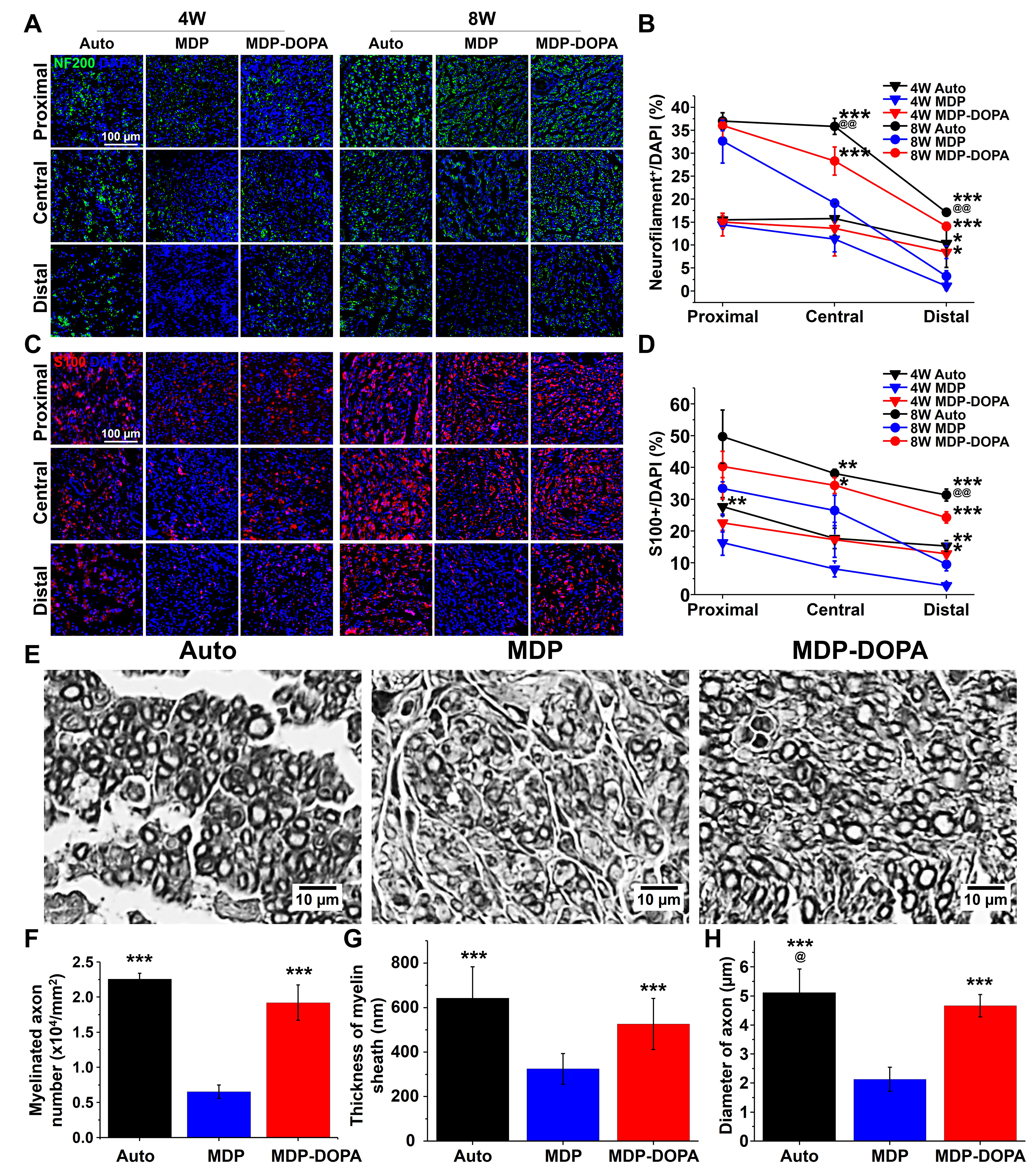
Figure 5. (A) Immunohistochemical evaluation of neurofilament 200 (NF200) expression at 4 and 8 weeks (scale bar = 100 μm); (B) Quantification of NF200 positive expression (*P < 0.05 and ***P < 0.001 vs. MDP, @@P < 0.01 vs. MDP-DOPA; statistical comparisons were conducted among groups at each time point); (C) Immunohistochemical evaluation of S100 at 4 and 8 weeks (Scale bar = 100 μm); (D) Quantification of S100 positive expression (*P < 0.05, **P < 0.01, and ***P < 0.001 vs. MDP; @@P < 0.01 vs. MDP-DOPA; statistical comparisons were conducted among groups at each time point); (E) Cross section of distal region of remyelinated nerve using osmic acid staining (Scale bar = 10 μm); Quantification results of (F) myelinated axon number, (G) thickness of myelin sheath, and (H) axon diameter (***P < 0.001 vs. MDP, @P < 0.05 vs. MDP-DOPA). All values denote the mean ± SD derived from four independent biological replicates. MDP: Milk-derived protein; DOPA: dihydroxyphenylalanine; SD: standard deviation; DAPI: 4′,6-diamidino-2-phenylindole.
Remyelination in the distal nerve segment was confirmed by osmic acid staining at 8 weeks [Figure 5E][39,40]. The number of myelinated axons was significantly higher in the MDP-DOPA group (1.92 ± 0.25 × 104 mm-2) than in the MDP group (0.65 ± 0.10 × 104 mm-2) and was comparable to the autograft group (2.26 ± 0.08 ×
Immunomodulatory response at the early stage of regeneration in vivo
To investigate the early-stage immune response, the injury site was analyzed 1 week after implantation [Figure 6][41]. Immunohistochemical analysis showed high expression of the M1-related macrophage marker CD86 in the MDP group (79.17% ± 7.95%), whereas expression was significantly lower in the autograft (22.63% ± 6.01%) and MDP-DOPA groups (36.54% ± 6.33%) [Figure 6A and B]. Conversely, expression of the M2-related macrophage marker CD206 was higher in the MDP-DOPA group (39.91% ± 7.12%) than in the MDP group (21.78% ± 4.62%) [Figure 6A and C].
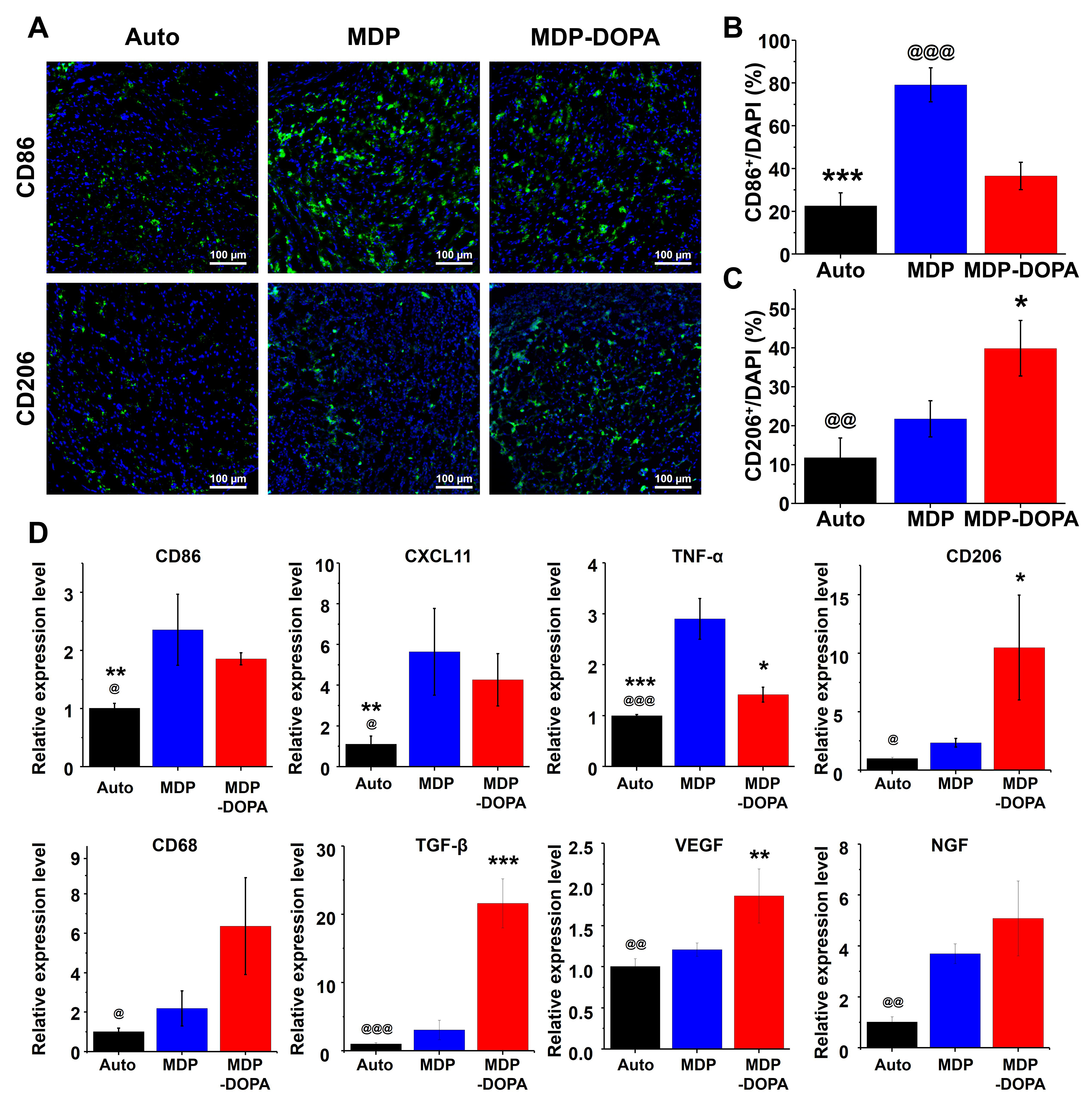
Figure 6. (A) Immunohistochemical staining of CD86 and CD206 at an early time point (scale bar = 100 μm); Quantification of positive expression of (B) CD86 and (C) CD206 (*P < 0.05 and ***P < 0.001 vs. MDP, @@P < 0.01 and @@@P < 0.001 vs. MDP-DOPA); (D) qRT-PCR evaluation of transcript levels for genes involved in inflammatory responses, macrophage polarization, and tissue repair calibrated against the autograft group (*P < 0.05, **P < 0.01, and ***P < 0.001 vs. MDP, @P < 0.05, @@P < 0.01, and @@@P < 0.001 vs. MDP-DOPA). Values represent mean ± SD from quadruplicate biological experiments. MDP: Milk-derived protein; DOPA: dihydroxyphenylalanine; qRT-PCR: quantitative reverse transcription polymerase chain reaction; SD: standard deviation; DAPI: 4′,6-diamidino-2-phenylindole.
qRT-PCR analysis of the remaining tissue region at 1 week confirmed these findings. M1-related markers (CD86, CXCL11, and TNF-α) showed significantly higher expression in the MDP and MDP-DOPA groups than in the autograft group[7,42]. By contrast, M2-related markers, especially CD206, TGF-β, and VEGF, showed significantly higher expression in the MDP-DOPA group than in both the autograft and MDP groups. Similarly, CD68 and NGF showed higher expression levels [Figure 6D].
Discussion
In this study, we developed multifunctional NGCs from MDP and PVA and functionalized them with DOPA to enhance peripheral nerve regeneration[43,44]. The fabrication process utilized a freeze–thaw method to create physical crosslinks, eliminating the need for chemical crosslinking agents that could cause denaturation and functional failure of bioactive proteins within casein.
The highly hydrophilic nature of PVA limits the adsorption of essential cell-binding proteins, thereby posing a challenge in providing cell-adhesion motifs[21]. To address this, we coated NGCs with DOPA, a mussel-derived adhesive protein[45]. This DOPA coating was hypothesized to enhance the adsorption of cell-binding proteins through electrostatic interactions, thereby promoting favorable cell responses such as the polarization of M2-lineage macrophages, which is critical for nerve regeneration[45]. Our characterization results confirmed that the DOPA coating process did not adversely affect the porous morphology or mechanical properties of the hydrogel, thereby ensuring that the conduit possesses the requisite stability to prevent luminal collapse after implantation[46,47]. Although their mechanical strength was lower than that of some brittle synthetic grafts, the flexibility and elongation capacity of our conduits were similar to those of human nerves, which, combined with the presence of bioactive peptides, may be more effective for nerve regeneration[30]. Because peripheral nerve repair is generally performed under tension-free coaptation, an extremely high tensile strength is not a prerequisite for successful implantation.
The nerve repair process after injury involves a complex immune response, starting with an inflammatory phase dominated by M1 macrophages, followed by a regenerative phase promoted by M2 macrophages[41,48]. We hypothesized that the bioactive peptides within the MDP and DOPA coatings would modulate this response to favor regeneration. Our in vitro results strongly supported this finding, demonstrating that the MDP-DOPA surface significantly promoted a shift from the M1 to the M2 macrophage phenotype. This shift can be attributed to the κ-casein fragments in MDP, which induce M2 polarization via TLR stimulation, and the enhanced cell adhesion provided by the DOPA surface, which facilitates interaction with macrophage integrins[15,16].
Although we acknowledge the inherent complexity and heterogeneity of macrophage subpopulations in vivo beyond this binary framework, our comprehensive cytokine array analysis of the macrophage secretome [Supplementary Figure 5] directly validated this pro-regenerative transition. Secretome profiling showed that the MDP-DOPA interface robustly suppressed a broad spectrum of pro-inflammatory cytokines, while concurrently driving elevated secretion of vital neurotrophic and angiogenic factors, establishing a highly favorable microenvironment for downstream tissue remodeling.
Furthermore, the secretome from M2-polarized macrophages promotes axonal regrowth[34,36,49]. Our study confirmed this indirectly, as CM from macrophages cultured on MDP-DOPA (MD-CM) significantly enhanced neurite outgrowth in PC12 cells to a level comparable to NDM. This potent neurogenic effect is likely a synergistic result of M2-secreted paracrine factors such as IL-10 and the direct action of bioactive casein peptides such as β-casomorphins and FLPYPY, released from the hydrogel. Importantly, PC12 cells maintained excellent viability and achieved highly accelerated neurite outgrowth under MD-CM conditions, demonstrating the exceptional cytocompatibility and the complete absence of cytotoxicity from our hydrogel platform and its degradation byproducts [Supplementary Figure 6].
The superior in vitro performance of MDP-DOPA translated into remarkable in vivo efficacy. In a rat sciatic nerve defect model, the MDP-DOPA conduit led to functional recovery (SFI and electrophysiology) and prevented muscle atrophy, significantly better than unmodified MDP and closely approaching the therapeutic trajectory of the clinical autograft gold standard. Although commercially available or well-established natural polymer scaffolds such as collagen- or chitosan-based conduits are frequently investigated as conventional controls, their clinical efficacy in large nerve gaps remains heavily constrained by poor mechanical stability, leading to luminal collapse and a lack of active immunomodulatory cues, often resulting in regenerative outcomes far inferior to autografts[3,4]. By contrast, our study used an autograft, the clinical gold standard for peripheral nerve repair, as the primary positive control to establish the most rigorous benchmark for functional integration. The fact that the soft, bioinstructive MDP-DOPA conduit achieved histological and functional recovery comparable to this ultimate clinical standard negates the necessity for an intermediate natural polymer control, further highlighting its superior potential for clinical translation. The MDP-DOPA group showed significantly greater axonal regrowth, Schwann cell migration, and remyelination (thicker myelin sheaths and larger axon diameters) in the distal nerve segment than the MDP group.
A key aspect of our proposed mechanism is early-stage immunomodulation at the injury site[6,39]. Analysis at one week post-implantation revealed that the MDP-DOPA conduit environment was dominated by M2 macrophages, in stark contrast to the M1-dominant environment in the MDP group. qRT-PCR data further confirmed that the tissue within the MDP-DOPA group showed high expression of M2-related cytokines and growth factors, including TGF-β, VEGF, and NGF. These factors are crucial for recruiting Schwann and endothelial cells, which in turn create the regenerative microenvironment necessary for robust axonal growth and functional recovery[6-8,41,42]. Thus, MDP-DOPA actively orchestrated the sequential processes of regeneration, beginning with modulation of the initial immune response. The predominant presence of M1 macrophages in the MDP group likely explains their inability to effectively transition to the healing phase, leading to poorer regenerative outcomes[33].
Study limitations
Despite the encouraging functional, histological, and immunomodulatory outcomes observed in this study, some limitations should be considered. First, the in vivo evaluation used a 10-mm sciatic nerve defect in adult male rats, with the longest observation period being limited to 8 weeks. This model was sufficient to demonstrate the regenerative efficacy of the MDP-DOPA conduit relative to the MDP conduit and autograft control. Longer follow-up periods and validation in more challenging nerve defects or larger animal models will be required to confirm the durability and broader translational applicability of the observed effects.
Second, we primarily evaluated degradation under controlled in vitro conditions using a collagenase-containing medium. The in vivo findings confirmed that the conduit maintained its tubular architecture without significant collapse at 8 weeks; however, the quantitative in vivo degradation kinetics, local tissue response to degradation residues, and long-term safety were not comprehensively assessed. Therefore, future studies should examine the temporal relationships among conduit degradation, tissue remodeling, and long-term biocompatibility.
Finally, although the present results support the involvement of MDP-derived bioactive components and DOPA-mediated cellular interactions in macrophage modulation and nerve regeneration, the exact primary peptide sequences and their specific downstream signaling cascades operating in this hydrogel platform were not individually isolated or blocked in the present study. While the observed M2 polarization and neurite outgrowth are consistent with the known bioactivities of κ-casein derivatives and β-casomorphins, future studies focused on peptide-specific sequence analysis and pathway inhibition will be necessary to further refine the underlying molecular mechanism. The current study characterized cumulative total protein release and changes in macrophage-associated gene and protein profiles, providing evidence for the proposed bioactivity; however, peptide-specific identification, quantitative release analysis, and pathway-blocking experiments are necessary to define the detailed molecular mechanisms. Additional studies would strengthen, rather than alter, the regenerative effects demonstrated in this study.
CONCLUSION
In this study, we fabricated a milk-derived casein NGC via a simple physical crosslinking process with PVA. The MDP surface was easily modified by immersion in a DOPA solution to promote interactions with the migrating host cells. The DOPA coating process did not affect surface deformation or mechanical properties. Seeding of RAW 264.7 cells onto MDP-DOPA promoted lineage commitment toward M2 macrophages due to immunomodulatory peptide fragments derived from κ-casein. In addition, MD-CM derived from RAW 264.7 cells cultured on MDP-DOPA improved neurogenic differentiation of PC12 cells via β-casomorphins and FLPYPY sequence peptides. In an in vivo animal study, MDP-DOPA induced remarkable nerve regeneration in a rat sciatic nerve injury model, as confirmed by SFI, electrophysiological, histological, IHC, and qRT-PCR analyses. Moreover, MDP-DOPA could induce an M2 phenotype macrophages and promote the secretion of nerve regeneration-related cytokines and proteins at the proximal injury site in the early stage. MDP-DOPA rapidly accelerated nerve regeneration via these bioactive peptides. Our unique MDP-DOPA scaffold could be used for various tissue-engineering applications without external stimulation.
Significance
Peripheral nerve repair remains challenging because the current NGCs lack the ability to actively modulate the immune microenvironment and support robust regeneration. In this study, we developed a milk-derived immunomodulatory hydrogel conduit functionalized with DOPA that promoted macrophage polarization toward a pro-regenerative phenotype and enhanced neurite outgrowth. In a rat sciatic nerve defect model, the conduit achieved functional and histological recovery comparable to autografts without in vivo delivery of exogenous growth factors. This study introduced a growth factor-free biomaterial strategy that integrates intrinsic bioactivity and immunomodulation, thereby providing a new design paradigm for next-generation NGCs and regenerative biomaterials.
DECLARATIONS
Authors’ contributions
Conceptualization, methodology, data curation, formal analysis, investigation, and writing - original draft: Jeon, J.
Methodology, data curation, formal analysis, and writing - original draft: Lee, M. S.; Kim, C.
Investigation and writing - review and editing: Park, J. H.; Chung, Y.; Yoon, J. K.; Ryu, H.
Investigation: Kim, E.
Supervision, funding acquisition, and writing - review and editing: Joung, Y. K.
Conceptualization, supervision, funding acquisition, writing - original draft, and writing - review and editing: Yang, H. S.
Availability of data and materials
Raw data and methods supporting the findings of this study are available in this Article and its Supplementary Materials. Additional data are available from the corresponding author upon request.
AI and AI-assisted tools statement
During the manuscript preparation, Google Gemini 3.1 Flash Image (Nano Banana 2; version 3.1, released February 26, 2026) was used to generate a background neuronal image in the graphical abstract. The authors created all other graphical elements and final compositions in Microsoft PowerPoint. The AI tool did not influence the study design, data collection, analysis, interpretation, or scientific content. All the authors take full responsibility for the accuracy, integrity, and final content of the manuscript.
Financial support and sponsorship
This work was supported by a National Research Foundation of Korea grant funded by the Korean government (MSIT) (2023R1A2C1006750), a Korean Fund for Regenerative Medicine (KFRM) grant funded by the Korean government (Ministry of Science and ICT, Ministry of Health & Welfare) (KFRM 25A0105L1), Basic Science Research Capacity Enhancement Project through Korea Basic Science Institute (National Research Facilities and Equipment Center) grant funded by the Ministry of Education (Grant No. 2019R1A6C1010033 and RS-2026-25539507), and the Parts Technology Development Program (2410017061, RS-2024-00434907) funded by the Ministry of Trade, Industry & Energy (MOTIE, Republic of Korea).
Conflicts of interest
Lee, M. S. is affiliated with R&D Center-ReCM BIO Co., while the other authors have declared that they have no conflicts of interest.
Ethical approval and consent to participate
All animal experimental procedures were reviewed and approved by the Institutional Animal Care and Use Committee (IACUC) at Dankook University (Approval No. DKU-22-001) and were performed in strict accordance with the NIH Guide for the Care and Use of Laboratory Animals and institutional guidelines.
Consent for publication
Not applicable.
Copyright
© The Author(s) 2026.
Supplementary Materials
REFERENCES
1. Liu, C.; Sun, M.; Lin, L.; et al. Potentially commercializable nerve guidance conduits for peripheral nerve injury: past, present, and future. Mater. Today. Bio. 2025, 31, 101503.
2. Shi, S.; Tang, J.; Lu, Y.; Xu, S.; Wang, M.; Wan, S. A burden of nerve injury from a global perspective, 1990-2021: an analysis of incidence, prevalence, and years lived with disability. Front. Neurol. 2025, 16, 1669662.
3. Maita, K. C.; Garcia, J. P.; Avila, F. R.; et al. Evaluation of the aging effect on peripheral nerve regeneration: a systematic review. J. Surg. Res. 2023, 288, 329-40.
4. Sarker, M.; Naghieh, S.; McInnes, A. D.; Schreyer, D. J.; Chen, X. Strategic design and fabrication of nerve guidance conduits for peripheral nerve regeneration. Biotechnol. J. 2018, 13, e1700635.
5. Redolfi Riva, E.; Özkan, M.; Contreras, E.; et al. Beyond the limiting gap length: peripheral nerve regeneration through implantable nerve guidance conduits. Biomater. Sci. 2024, 12, 1371-404.
6. Liu, P.; Peng, J.; Han, G. H.; et al. Role of macrophages in peripheral nerve injury and repair. Neural. Regen. Res. 2019, 14, 1335-42.
7. Gensel, J. C.; Zhang, B. Macrophage activation and its role in repair and pathology after spinal cord injury. Brain. Res. 2015, 1619, 1-11.
8. Wu, G.; Wen, X.; Kuang, R.; et al. Roles of macrophages and their interactions with schwann cells after peripheral nerve injury. Cell. Mol. Neurobiol. 2023, 44, 11.
9. Rios, R.; Jablonka-Shariff, A.; Broberg, C.; Snyder-Warwick, A. K. Macrophage roles in peripheral nervous system injury and pathology: allies in neuromuscular junction recovery. Mol. Cell. Neurosci. 2021, 111, 103590.
10. Xu, H.; Yu, Y.; Zhang, L.; et al. Sustainable release of nerve growth factor for peripheral nerve regeneration using nerve conduits laden with bioconjugated hyaluronic acid-chitosan hydrogel. Compos. Part. B. Eng. 2022, 230, 109509.
11. Fioranelli, M.; Roccia, M. G. Twenty-five years of studies and trials for the therapeutic application of IL-10 immunomodulating properties. From high doses administration to low dose medicine new paradigm. J. Integr. Cardiol. 2014, 1, 2-6.
12. Yao, X.; Xue, T.; Chen, B.; et al. Advances in biomaterial-based tissue engineering for peripheral nerve injury repair. Bioact. Mater. 2025, 46, 150-72.
13. Nielsen, S. D.; Liang, N.; Rathish, H.; et al. Bioactive milk peptides: an updated comprehensive overview and database. Crit. Rev. Food. Sci. Nutr. 2024, 64, 11510-29.
14. Trompette, A.; Claustre, J.; Caillon, F.; Jourdan, G.; Chayvialle, J. A.; Plaisancie, P. Milk bioactive peptides and β-casomorphins induce mucus release in rat jejunum. J. Nutr. 2003, 133, 3499-503.
15. Kitazawa, H.; Yonezawa, K.; Tohno, M.; et al. Enzymatic digestion of the milk protein beta-casein releases potent chemotactic peptide(s) for monocytes and macrophages. Int. Immunopharmacol. 2007, 7, 1150-9.
16. Lalor, R.; O’Neill, S. Bovine κ-casein fragment induces hypo-responsive M2-like macrophage phenotype. Nutrients 2019, 11, 1688.
17. Sakaguchi, M.; Ishikawa, C.; Nishimura, T.; Sugihara, E.; Matsumura, E. Neurite outgrowth-stimulating peptide derived from bovine kappa-casein. Biosci. Biotechnol. Biochem. 2004, 68, 2653-5.
18. Hsiang, S. W.; Tsai, C. C.; Tsai, F. J.; Ho, T. Y.; Yao, C. H.; Chen, Y. S. Novel use of biodegradable casein conduits for guided peripheral nerve regeneration. J. R. Soc. Interface. 2011, 8, 1622-34.
19. Omidian, H.; Dey Chowdhury, S.; Babanejad, N. Cryogels: advancing biomaterials for transformative biomedical applications. Pharmaceutics 2023, 15, 1836.
20. Adelnia, H.; Ensandoost, R.; Shebbrin Moonshi, S.; Gavgani, J. N.; Vasafi, E. I.; Ta, H. T. Freeze/thawed polyvinyl alcohol hydrogels: present, past and future. Eur. Polym. J. 2022, 164, 110974.
21. Ino, J. M.; Chevallier, P.; Letourneur, D.; Mantovani, D.; Le Visage, C. Plasma functionalization of poly(vinyl alcohol) hydrogel for cell adhesion enhancement. Biomatter 2013, 3, e25414.
22. Cheon, S. Y.; Kim, J.; Lee, K. Y.; Lee, C. M. Poly (l-lactic acid) membrane crosslinked with Genipin for guided bone regeneration. Int. J. Biol. Macromol. 2021, 191, 1228-39.
23. Lee, M. S.; Jeon, J.; Park, S.; Lim, J.; Yang, H. S. Rationally designed bioactive milk-derived protein scaffolds enhanced new bone formation. Bioact. Mater. 2023, 20, 368-80.
24. Porzionato, A.; Barbon, S.; Stocco, E.; et al. Development of oxidized polyvinyl alcohol-based nerve conduits coupled with the ciliary neurotrophic factor. Materials 2019, 12, 1996.
25. Kim, S. H.; Lee, J. H.; Hyun, H.; et al. Near-infrared fluorescence imaging for noninvasive trafficking of scaffold degradation. Sci. Rep. 2013, 3, 1198.
26. Kim, S. M.; Lee, M. S.; Jeon, J.; et al. Biodegradable nerve guidance conduit with microporous and micropatterned poly(lactic-co-glycolic acid)-accelerated sciatic nerve regeneration. Macromol. Biosci. 2018, 18, e1800290.
27. Kim, S. J.; Park, W. H. Polydopamine- and polyDOPA-coated electrospun poly(vinyl alcohol) nanofibrous membranes for cationic dye removal. Polym. Test. 2020, 89, 106627.
28. Arakawa, C.; Gunnarsson, C.; Howard, C.; et al. Biophysical and biomolecular interactions of malaria-infected erythrocytes in engineered human capillaries. Sci. Adv. 2020, 6, eaay7243.
29. Decarli, M. C.; Amaral, R.; Santos, D. P. D.; et al. Cell spheroids as a versatile research platform: formation mechanisms, high throughput production, characterization and applications. Biofabrication 2021, 13, 032002.
30. Clements, B. A.; Bushman, J.; Murthy, N. S.; Ezra, M.; Pastore, C. M.; Kohn, J. Design of barrier coatings on kink-resistant peripheral nerve conduits. J. Tissue. Eng. 2016, 7, 2041731416629471.
31. Wan, T.; Wang, Y. L.; Zhang, F. S.; et al. The porous structure of peripheral nerve guidance conduits: features, fabrication, and implications for peripheral nerve regeneration. Int. J. Mol. Sci. 2023, 24, 14132.
32. McQuillan, T. J.; Zelenski, N. A.; Ghareeb, P. A. The role of nerve tension on nerve repair success. J. Hand. Surg. Glob. Online. 2024, 6, 691-3.
33. Li, M.; Wang, X.; Qi, B.; et al. Treatment of syringomyelia characterized by focal dilatation of the central canal using mesenchymal stem cells and neural stem cells. Tissue. Eng. Regen. Med. 2024, 21, 625-39.
34. Kigerl, K. A.; Gensel, J. C.; Ankeny, D. P.; Alexander, J. K.; Donnelly, D. J.; Popovich, P. G. Identification of two distinct macrophage subsets with divergent effects causing either neurotoxicity or regeneration in the injured mouse spinal cord. J. Neurosci. 2009, 29, 13435-44.
35. Hu, X.; Leak, R. K.; Shi, Y.; et al. Microglial and macrophage polarization - new prospects for brain repair. Nat. Rev. Neurol. 2015, 11, 56-64.
36. Zigmond, R. E.; Echevarria, F. D. Macrophage biology in the peripheral nervous system after injury. Prog. Neurobiol. 2019, 173, 102-21.
37. Baehr, L. M.; Hughes, D. C.; Lynch, S. A.; et al. Identification of the MuRF1 skeletal muscle ubiquitylome through quantitative proteomics. Function 2021, 2, zqab029.
38. Wu, J.; Han, Q.; Gui, D.; Qian, Y. Multidimensional advances in neural interface technology for peripheral nerve repair: from material innovation to clinical translation. Mater. Today. Bio. 2025, 34, 102092.
39. He, X.; Yu, Y.; Awatramani, R.; Lu, Q. R. Unwrapping myelination by microRNAs. Neuroscientist 2012, 18, 45-55.
40. Yuan, Y. S.; Niu, S. P.; Yu, Y. L.; et al. Reinnervation of spinal cord anterior horn cells after median nerve repair using transposition with other nerves. Neural. Regen. Res. 2019, 14, 699-705.
41. Yum, Y.; Park, S.; Nam, Y. H.; et al. Therapeutic effect of schwann cell-like cells differentiated from human tonsil-derived mesenchymal stem cells on diabetic neuropathy in db/db mice. Tissue. Eng. Regen. Med. 2024, 21, 761-76.
42. Rotshenker, S. Wallerian degeneration: the innate-immune response to traumatic nerve injury. J. Neuroinflammation. 2011, 8, 109.
43. Memic, A.; Colombani, T.; Eggermont, L. J.; et al. Latest advances in cryogel technology for biomedical applications. Adv. Ther. 2019, 2, 1800114.
44. Feng, C.; Chen, M.; Qu, D. H. Physically cross-linked hydrogels based on poly(vinyl alcohol) and gelatin for independent modulation of mechanical cues in cell adhesion studies. ACS. Appl. Bio. Mater. 2026, 9, 3029-37.
45. Ma, T.; Wang, C. X.; Ge, X. Y.; Zhang, Y. Applications of polydopamine in implant surface modification. Macromol. Biosci. 2023, 23, e2300067.
47. Tian, Y.; Li, Y.; Xu, M.; Li, W.; Cong, H.; Yu, B. Polyvinyl alcohol hydrogels: structure, preparation, modification strategies and biomedical applications. Eur. Polym. J. 2026, 244, 114517.
48. Gopalakrishnan, B.; Galili, U.; Saenger, M.; et al. α-Gal nanoparticles in CNS trauma: II. Immunomodulation following spinal cord injury (SCI) improves functional outcomes. Tissue. Eng. Regen. Med. 2024, 21, 437-53.
Cite This Article
 Open Access
How to Cite
Jeon, J.; Lee, M. S.; Kim, C.; Park, J. H.; Chung, Y.; Kim, E.; Yoon, J.K.; Ryu, H.; Joung, Y. K.; Yang, H. S. Flexible and immunomodulatory milk-derived protein hydrogels as transient interfacing scaffolds for peripheral nerve integration. Soft Sci. 2026, 6, 86. https://dx.doi.org/10.20517/ss.2026.137
Download Citation
If you have the appropriate software installed, you can download article citation data to the citation manager of your choice. Simply select your manager software from the list below and click on download.
Export Citation File
Type of Import
Tips on Downloading Citation
Citation Manager File Format
Type of Import
Direct Import: When the Direct Import option is selected (the default state), a dialogue box will give you the option to Save or Open the downloaded citation data. Choosing Open will either launch your citation manager or give you a choice of applications with which to use the metadata. The Save option saves the file locally for later use.
Indirect Import: When the Indirect Import option is selected, the metadata is displayed and may be copied and pasted as needed.
About This Article
Special Topic
Copyright

Data & Comments
Data
0












Comments
Comments must be written in English. Spam, offensive content, impersonation, and private information will not be permitted. If any comment is reported and identified as inappropriate content by OAE staff, the comment will be removed without notice. If you have any queries or need any help, please contact us at support@oaepublish.com.