


fig1
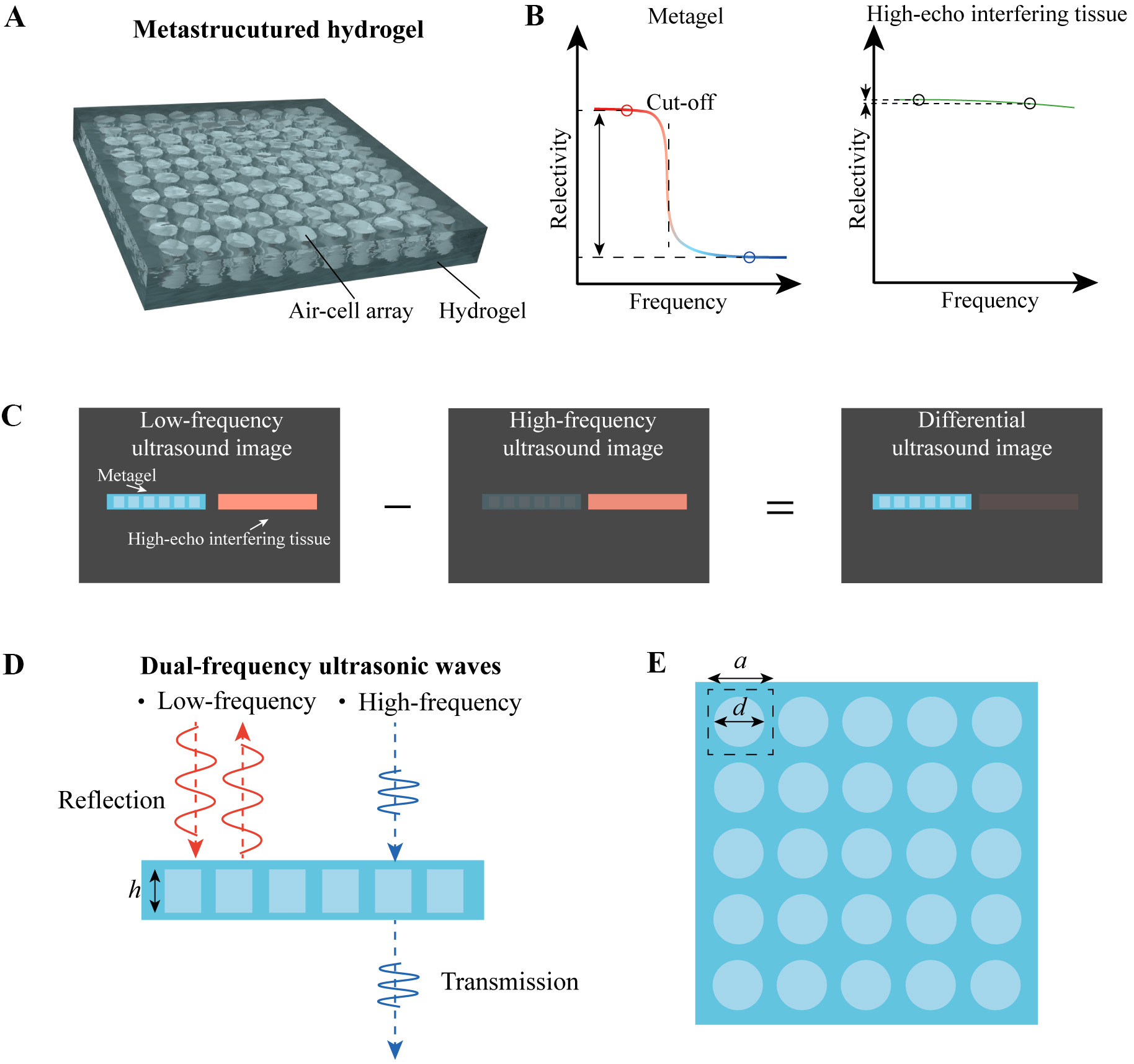
Figure 1. Schematic of the metagel film. (A) Schematic of the metagel film with periodically arranged air-cells within the hydrogel; (B) Schematic of the metagel reflection spectrum, showing a steep decrease in reflectivity at the cutoff frequency and a significant contrast across high and low frequency ranges; (C) Schematic of differential imaging, which achieves high contrast to eliminate interference from high-echo tissues; (D) Schematic of dual-frequency ultrasound excitation mode, in which low-frequency ultrasound is reflected while high-frequency ultrasound is transmitted. The height of the air-cell scatterer is denoted as h; (E) Geometric parameters influencing metagel performance, including the diameter d of the air-cell scatterer and the lattice constant a.


